CHAPTER 16Treatment of Fungal Endometritis
Treating equine fungal endometritis can be a frustrating endeavor to undertake. Oftentimes mares become reinfected or remain infected with fungi, are culture positive with a bacterial endometritis the next cycle, or are classified as susceptible to infection/uterine fluid accumulation. Generally, mares affected are 10+-years-old with a history of infertility. Most treatments for fungal endometritis have been extrapolated from other species or from other disease processes. Specific controlled intrauterine fungal treatment studies are lacking in the horse. The information presented in this chapter is based on what is available in the literature, the author’s experience, and information from colleagues. This chapter will be broken down into a description of fungi, fungal endometritis, and the diagnosis and treatment of fungal endometritis. By following a systematic approach with a longer duration of therapy, infection can be eliminated in many mares, allowing for subsequent breeding.
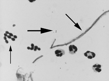
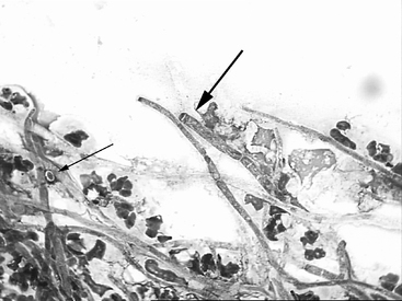
Fungi are classified either as single-celled organisms, called yeast, or multi-cellular filamentous organisms, called molds. The filamentous cells within molds are called hyphae. Hyphae may also form tangled masses called mycelia. Cytological specimens may demonstrate yeast or hyphae (Figures 16-1 and 16-2), whereas mycelia are usually identified in fluid from a uterine lavage (Figure 16-3). Yeasts are typically more suited to growth in liquids, whereas hyphae are better adapted for penetrating solid substrates. Thus, when hyphae are identified on cytology or biopsy, thought should be directed toward treatment of a more deep seated infection within the endometrium. Yeasts are 3 to 5 μm round-to-oval single-cell organisms and often have a capsule surrounding them that excludes dyes (Figure 16-2). By comparison, neutrophils are typically 10 μm in diameter.
A number of different fungi have been isolated from the mare’s reproductive tract, with Candida spp and Aspergillus spp identified most commonly. Table 16-1 lists the fungi that have been isolated from the mare’s reproductive tract. Certain fungi, such as Candida spp, may normally be present as commensal organisms in the gastrointestinal tract, vagina, or oral mucosa. Candida may be present as both yeast cells and hyphal elements on cytology. Candida albicans is able to adhere to vaginal epithelium better than other Candida spp; thus it is isolated from the majority of Candida infections. Estrogen enhances the ability of Candida to adhere to vaginal tissue; consequently, drug therapy with estrogens may increase the risk for this fungal infection. Nystatin, clotrimazole, ketoconazole and amphotericin B have been reported as effective against Candida spp. Some of the newer azole drugs, such as fluconazole, may also be effective. In contrast to Candida, Aspergillus spp are in hyphal form and may exhibit conidiophores. Aspergillus has been reported to invade tissues, making topical therapy less effective. Macrophages are particularly effective against Aspergillis, and when macrophage function is decreased, tissue invasion is more likely. Hyphal forms are too large for phagocytosis by polymorphonuclear cells; consequently, they must be damaged by substances released by polymorphonuclear leukocytes and mononuclear phagocytes. Amphotericin B is often employed in the treatment of Aspergillis infections, although the azole compounds should also be effective, as described later.
Table 16-1 Fungi that have Been Reported in Isolates from the Associated Form


