Picture 14.1
1 aorta abdominalis, 2 arteria mesenterica cranialis, 3 arteria celiaca, 4 arteria renalis lat. sin., 5 truncus lumbalis, 6 truncus intestinalis, 7 lymphatic collector of left kidney, 8 cysterna chyli, 9 ductus thoracicus. *The area where aorta was interrupted and eventually the thoracic duct at organ removal (see Chap. 13, small bowel transplantation )
14.3 Lymph Vessels Graft Procurement
Animals
The experiment was carried out on inbred males of strain Brown-Norway (BN) as donors and inbred males Lewis (LEW) as recipients.
Equipment + Material
Scissors, forceps, micro-scissors (straight, curved), two micro-forceps (straight, curved), micro-needle holder, Operating microscope, 9–0 nylon suture, 7–0 and 4–0 silk suture, cotton swabs, gauze, saline solution, heparin solution, heating pad, needles, syringes.
14.3.1 Operation of Donor
Operation of donor differs from isolated small bowel transplantation in some steps. The initial phase of operation, position of animal, anesthesia and instruments are identical with the description in Chap. 13. After release of aortic caudal part above the bifurcation, and after release of portal vein and both ends of jejunum, start with operation of the lymphatic vessel s .
1.
Gently separate the aorta from the retroperitoneum up to the base of arteria mesenterica cranialis
2.
Between the ligatures, look up and cut up both trunci lumbales and the lymphatic collector of the left kidney
3.
Dissect the ductus thoracicus from animal’s right side (Picture 14.2). Follow the ligation of the right renal artery which deviates from the aorta on the same level as the arteria mesenterica cranialis and lymphatic collector of the right kidney
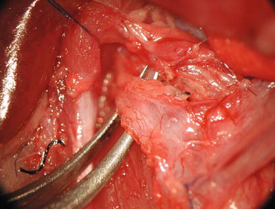
Picture 14.2
Detail of thoracic duct
4.
Release the aorta from the mesentery up to above the base of the celiac artery. Be extremely careful at this point in order to avoid damage of the cysterna chyli, which is attached to the dorsal side of the aorta in the place of the base of mesenteric artery and the thoracic duct on the dorsolateral side of the aorta
5.
Above the base of the celiac artery, separate the thoracic duct from the aorta and continue the blunt dissection of thoracic duct from the aorta and continue up to diaphragm. Ligate the small lumbar arteries coming out of the lumbar aorta
6.
Ligate the celiac artery together with the connecting lymphatic vessel s
7.
After blunt dissection and separation of the left part of aortal segment from retro-peritoneum, the graft is ready to be rinsed with preservative solution. The graft consists of a 7–10 cm long segment of jejunum with the corresponding part of the mesentery and portal vein, aortal segment from bifurcation up to the base of the celiac artery and the lymphatic vessel s collecting lymph from the small bowel graft and opening out into the thoracic duct (Picture 14.3)
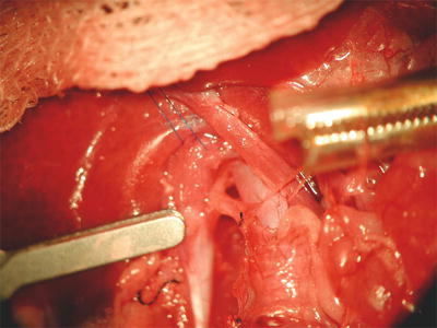
Picture 14.3
Situation before rinsing the graft with preservative solution, separated aorta, portal vein and thoracic duct
8.
Ligate the aorta above the bifurcation and above the celiac artery base
9.
Insert the cannula for perfusion into the aorta just above the bifurcation
10.
Interrupt the portal vein before the insertion into liver and start with perfusion with approximately 3–4 ml of physiological saline solution with heparin
11.
After the perfusion is finished, interrupt the aorta in the site of cannula insertion and above the ligature of the aorta below the diaphragm
12.
Remove the graft and place it into the cold saline solution
13.


Euthanize the animal
Stay updated, free articles. Join our Telegram channel
Full access? Get Clinical Tree
