Figure 26.2 Computed tomography image of an alpaca with extensive bony proliferation surrounding the second molar tooth of the right mandible, loss of alveolar bone, evidence of osteomyelitis, and disruption of the tooth.
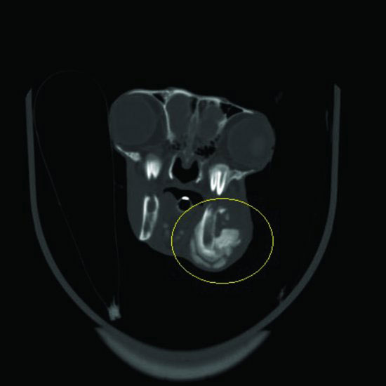
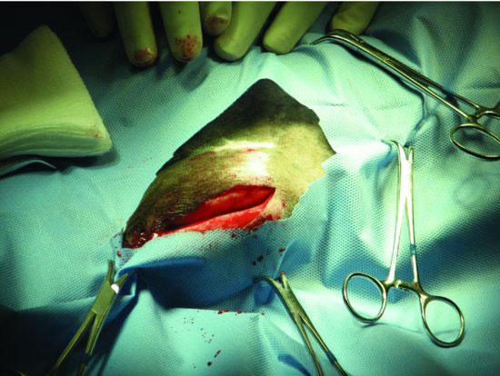
Figure 26.4 Myoperiosteal elevation flap reflected dorsally to expose the underlying bone to provide access to surgically approach the lateral alveolar plate and entry into the oral cavity to exposure the crown of the effected tooth.
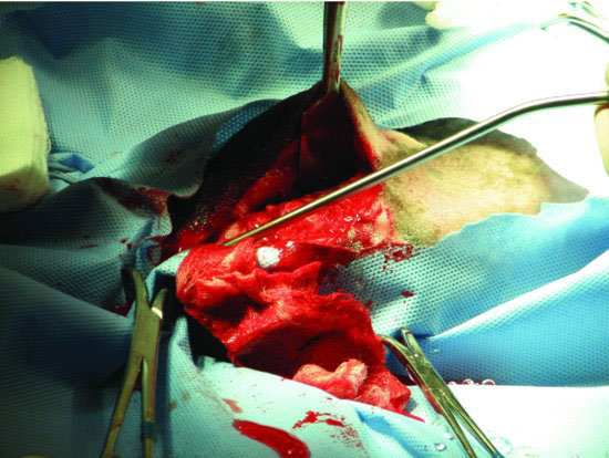
Figure 26.5 The lateral alveolar plate of bone overlying the effected tooth is most easily removed using a rotating burr such as this pneumatic osteotome.
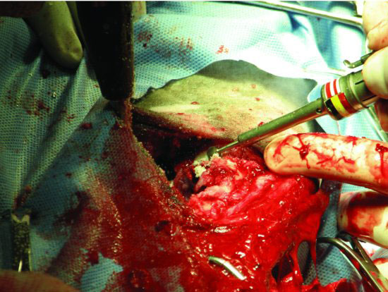
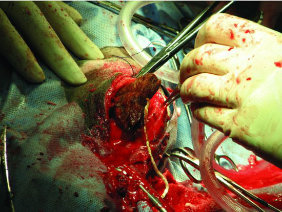
Figure 26.6 After tooth extraction, a temporary plug may be inserted using silicone dental putty, polymethylmethacrylate, or gauze sponges (depicted here).
Stay updated, free articles. Join our Telegram channel

Full access? Get Clinical Tree


