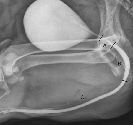
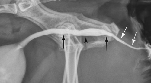
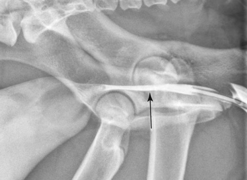
The urethra is a tubular organ that serves as an outlet for urine from the urinary bladder and, in the male, semen and reproductive secretions. Anatomically, the male urethra comprises three major areas: the prostatic urethra, membranous urethra, and penile urethra (Fig. 40-1).1 Together, the prostatic and membranous portions make up the pelvic part of the urethra.2 The prostatic urethra is surrounded completely by the prostate gland in the dog, although only dorsolaterally in the cat.3 The membranous urethra is enveloped by cavernous tissue and the urethralis muscle.3 The penile urethra extends from the ischial arch to the tip of the penis. This part of the urethra is also known as the cavernous urethra because the majority of it is encased by erectile tissues of the penis.2 The most distal part of the penile urethra is received and protected by the ventral urethral groove of the os penis. The arrangement of urethral muscle and surrounding spongy tissues confers a diffuse sphincter mechanism to the urethra in male dogs. The female urethra is comparatively shorter and wider than in males and entirely pelvic in position. The external urethral orifice lies caudal to the vestibulovaginal junction, where a muscular urethral sphincter is present.2 In the cat, the urinary bladder is typically intraabdominal in position, and a relatively long prepelvic part of the urethra extends between the neck of the urinary bladder and the pelvic urethra.3 Retrograde urethrography is a technique to examine the urethra using positive-contrast medium. Only water-soluble, iodinated contrast medium should be used. The contrast medium can be diluted with sterile saline to a final concentration of 15% (150 mg iodine [I]/mL).1,4 Patients generally require sedation, and evacuation of the colon and rectum through defecation or cleansing enemas is recommended. A fully distended urinary bladder provides counterpressure to contrast-medium injection to allow maximal distention of the urethra.5 Often cystography is performed initially, and distention of the bladder with contrast medium should be maintained for urethrography. If necessary, sterile saline can be infused into an inadequately distended bladder. In addition to the aforementioned radiographic projections, right ventral-left dorsal and left ventral-right dorsal views should be included because the standard ventrodorsal projection may be less informative owing to superimposition of the pelvic and penile urethra. In male dogs and cats, the procedure is performed by inserting a catheter into the distal penile urethra. A balloon-tipped (Foley) catheter is preferred in dogs, although a red rubber catheter may also be used.4,6 The bulb of the Foley catheter is gently inflated and left inflated no more than 15 minutes, because excessive and prolonged pressure can damage the urethra.7 In male cats, a red rubber or Tomcat catheter may be used (Fig. 40-2). Depending on patient size, a 5 to 20 mL injection of contrast medium is given and radiographs acquired during the delivery of the final 1 to 2 mL of the injection.4 The normal urethra should be smoothly margined along its entire length. The prostatic urethra is wider than the membranous urethra, although each can be variable in diameter depending on the degree of distention.8 The penile urethra has a generally uniform diameter in dogs; in cats, it is relatively narrow.9 Luminal narrowing with tapered margins may be seen normally at the ischial arch in dogs.4 Retrograde urethrography is challenging in female dogs and cats because of the relatively shorter urethra. A Foley catheter is inserted into the distal urethra and inflated gently. A 5 to 10 mL injection of contrast medium is used to distend the urethra and radiographs made at the end of injection (Fig. 40-3).4 Vaginocystourethrography is an alternate technique for evaluation of the female urethra and may be performed in dogs and cats (Fig. 40-4). Video 40-1 can be found on the accompanying Evolve website. General anesthesia is required when performing vaginocystourethrography. A Foley catheter is inserted into the vestibule and inflated. The tip of the catheter is removed from the bulb to avoid the tip entering the vagina and causing rupture on injection.10 Tissue forceps may be used to clamp the vulva to prevent expulsion of the catheter and leakage of contrast medium. Contrast medium at a volume of 1 mL/kg is injected slowly, avoiding high pressure. Contrast medium fills the vagina, and with increased pressure during injection, there is retrograde filling of the urethra. The normal urethra should be smoothly margined, and longitudinal striations attributable to mucosal folding may be seen depending on the degree of distention.4
The Urethra
Anatomy
Radiography and Urethrography
The Urethra