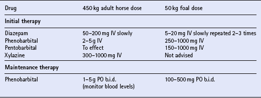
The nervous system

Chapter 17


The nervous system


Chapter contents


INTRODUCTION


CONGENITAL AND DEVELOPMENTAL PROBLEMS


INFLAMMATORY AND TOXICO-INFECTIOUS DISEASES


METABOLIC DISORDERS


TOXIC CONDITIONS


SPACE-OCCUPYING LESIONS


TRAUMA


POST-ANESTHETIC CENTRAL NERVOUS SYSTEM DISORDERS


CONDITIONS OF THE LOWER MOTOR NEURON UNIT


NEUROLOGIC ASPECTS OF “VICES”


NARCOLEPSY/CATALEPSY


INTRACAROTID INJECTION


GRASS SICKNESS (EQUINE DYSAUTONOMIA)


INTRODUCTION


With the advent of equine protozoal myeloencephalitis (q.v.) and West Nile virus (q.v.), neurologic diseases of horses have become increasingly significant worldwide. Since the laboratory diagnosis of those two diseases is not straightforward, and other diseases need disparate treatments and offer a different prognosis, there is increasing pressure on clinicians to make the correct clinical diagnosis.


In the neurologic examination of the equine patient, it is important to remember that the objective is to make an “anatomic diagnosis”, i.e. establish in which neurologic structures the lesion(s) are likely to be in order to result in the observed neurologic signs.


Mentation, cranial nerve reflexes, cutaneous reflexes, tail and anus function and gait should be carefully examined. Is the lesion central (i.e. brain or spinal cord) or in the lower motor neuron unit (lower motor neuron cell body, peripheral nerve, neuromuscular junction or muscle)? Can the signs be explained by a focal lesion or are they likely to be due to a multifocal or diffuse disease? The answer to those questions will dramatically narrow down the possible differential diagnoses and will determine the choice of diagnostic tests.


This chapter aims to provide the equine practitioner with a synopsis of the clinical findings, diagnostic tests, treatment and prognoses for common neurologic disorders.


CONGENITAL AND DEVELOPMENTAL PROBLEMS


HYDROCEPHALUS


The congenital form of hydrocephalus is rare in newborn foals. When it does occur, it is usually due to cerebral malformation and accumulation of fluid in the ventricles. The head may or may not be grossly enlarged or dome shaped. Most foals with congenital hydrocephalus die shortly after birth but cases can be diagnosed later as incidental findings. Severe dystocia (q.v.) may be associated with the condition if the head is grossly deformed.


Acquired hydrocephalus is also occasionally observed and is usually due to adhesions secondary to bacterial meningitis or intracranial hemorrhage. Adhesions obstruct the normal outflow of cerebrospinal fluid (CSF). The head is sometimes, but not always, grossly enlarged. Foals with acquired hydrocephalus may be obtunded. As intracranial CSF pressures increase, seizures (q.v.) may ensue.


CSF analysis in congenital hydrocephalus is usually normal. In acquired cases, it may reflect the primary disease. In foals, the fontanelles of the cranium are not open, thus ultrasonography is not useful. Computed tomography or magnetic resonance imaging is potentially the best means to obtain an ante mortem diagnosis.


There is no practical form of treatment with congenital or acquired hydrocephalus. The prognosis for long-term survival is poor.


CEREBELLAR ABIOTROPHY


Cerebellar abiotrophy is suspected to be an inherited disorder with an 8% incidence in some Arabian family lines. It is also considered an inherited disorder in Oldenburg light-breed horses and in Eriskay and Gotland ponies. Either gender can be affected. The diagnosis is based on breed, history and clinical signs. The signs may be present at birth, but can be difficult to recognize in foals <1 mo of age. The clinical signs are characterized by jerky head movements and hypermetria/dysmetria. Affected foals assume a wide-based stance at rest and may fall over backwards if asked to back up or elevate the head. As the foal ages, it will appear strong but ataxic and continue to exhibit a rhythmic head bobbing or head tremors, characteristic of cerebellar disease (q.v.). Blindness and gait paresis are not features of this disease.


There are no ante mortem tests. The CSF is normal. There is no treatment and affected foals should be euthanased because they may injure themselves or their handlers. Cerebellar abiotrophy is confirmed at post mortem by a cerebellum that weighs <10% of the weight of the whole brain, histologic evidence of thinning of the granular and molecular layers of the cerebellar cortex and degeneration of the Purkinje cells.


JUVENILE EPILEPSY OF FOALS


Juvenile epilepsy in foals is probably not a form of true inherited epilepsy. It is speculated to be a type of pediatric seizure that reflects the neonate’s low seizure threshold in response to temporary derangements. The temporary derangements can be caused by toxic, metabolic, physical, infectious or circulatory disorders.


A form of idiopathic epilepsy is occasionally reported in suckling and weanling foals, in particular those of the Arabian breed. Seizures begin between 1 and 12 mo of age. Several seizures may occur immediately prior to presentation. The seizures are characterized by episodes of sudden loss of consciousness with or without tonic-clonic activity. Affected foals can experience temporary central blindness (which can last a few hours to a few days). Head trauma due to thrashing is a common complication. Seizures do not usually persist into adulthood.


Once other causes for seizures have been ruled out, a diagnosis of idiopathic epilepsy can be given. Although the disease is self-limiting with age, seizures must be kept under control ( Table 17.1) to avoid permanent brain damage or head trauma. Initial anticonvulsant therapy with diazepam (5–20 mg IV) repeated two or three times will usually control the seizures. For maintenance anticonvulsant therapy, affected foals should be kept on phenobarbital (100–500 mg PO s.i.d. or b.i.d.) for a minimum of 10–14 days, or as long as is needed to avoid recurrence of seizure activity. Treatment with phenobarbital should be dose adjusted so that seizures can be controlled but the foal not overly sedated. Monitoring blood levels of the drug is advised and the non-toxic therapeutic range is approximately 5–30 μg/mL. The lowest dose that controls seizures should be used, as phenobarbital may sedate the foal and discourage nursing. The dose must be tapered down over 2 weeks before being discontinued, as abrupt discontinuation may precipitate seizures.


Pediatric epilepsy generally subsides as the foal ages, as long as seizure-related brain damage has not occurred. If blindness persists in the postictal phase, vision usually returns within a few days after seizure activity is controlled. The incidence of recurrence after puberty is unknown.


CERVICAL VERTEBRAL MALFORMATION


Cervical vertebral malformation (CVM) describes a group of malformations and malarticulation abnormalities of the cervical vertebrae of horses. It is a common cause of ataxia (q.v.) in horses all over the world and affects any breed of horse, but particularly Thoroughbreds. The disease stems from bone and joint malformations and is seen as two broad categories. Younger horses and foals are predisposed to deformation of the vertebral bodies with malarticulation and subluxation on flexion, while the spinal cord compression in older horses is generally due to osteoarthritis of the caudal articular processes.


The disease in young horses may be a form of a developmental orthopedic disease, i.e. related to rapid skeletal growth in genetically predisposed horses. Cervical vertebrae in those animals have lesions of osteochondrosis and epiphysitis similar to those seen in the limbs. Familial predisposition, high dietary energy intake and trauma to the neck probably all play a role, as young, large, fast-growing animals (often males) are affected.


An accident or injury to the neck is sometimes in the history, and clinical signs may have an abrupt onset. Mild to moderately affected horses show symmetrical ataxia with circumduction of the pelvic limbs, especially when the animal is made to walk in small circles. Proprioceptive deficits, toe dragging, dysmetria and varying degrees of upper motor neuron weakness (assessed by pulling on the horse’s tail at a walk) are also present. Thoracic limb deficits are usually less severe than pelvic limb deficits. The “slap test” for laryngeal adduction (q.v.) may be absent but there are no cranial nerve deficits. Neck pain or stiffness may be present, particularly in older horses.


The neurologic examination and plain radiographs of the vertebrae (C1–T2), while the horse is standing with the neck in a neutral position, are the most important diagnostic aids. In younger horses plain film radiographs reveal stenosis of the vertebral canal, often at C3–4 or C4–5. A corrected minimum sagittal diameter (the ratio of the absolute minimum sagittal diameter of the vertebral canal to the sagittal width of the vertebral body) of <0.5 is highly suggestive of a compressive lesion at C3–4 or C4–5.


Enlarged caudal epiphyses, a caudal extension of the dorsal aspect of the vertebral arch across the ventral articulation site of adjacent vertebrae, vertebral malalignment and degenerative/osteochondrosis-like changes in the articular processes often coexist. Greatly enlarged articular processes, with osteophytes, associated with hypertrophy and fibrosis of synovial membranes and joint capsule, can cause acute onset compression in older horses. Myelography, a technique used to assess spinal cord compression by the injection of radiodense contrast medium into the subarachnoid space, may be useful in determining the extent of cord compression and should be performed on horses in which surgical decompression and/or vertebral fusion are being considered.


In a research setting, the radiographic and neurologic deficits may be reversed in young (≤1 yr of age) animals by confinement (to limit further injury to the cord) and a severely protein- and energy-restricted diet. On the whole, however, the prognosis for return to athletic function of horses with CVM is poor. Affected horses may become so ataxic that they become a danger to themselves and horse handlers. Humane destruction is then advised. Surgical fusion of the vertebrae has been used to treat, and may improve, affected horses, but residual deficits often persist.


EQUINE DEGENERATIVE MYELOENCEPHALOPATHY


Equine degenerative myeloencephalopathy (EDM) is a disease of young horses of any breed, although a familial tendency has been observed in certain Appaloosa, Standardbred and possibly Morgan lines. The disease is thought to be related to vitamin E deficiency associated with lack of green forage, or the feeding of heat-processed pelleted rations. A history of vitamin E deficiency is, however, not evident in all cases. Neuroaxonal dystrophy in the brainstem and spinal cord results in gait abnormalities.


EDM is rarely seen in horses ≥4 yr of age. Symmetrical ataxia, hypometria and paresis usually appear in animals ≤6 mo of age. The disease is progressive, but the clinical signs have generally stabilized after the animal reaches 6–12 mo of age. Signs may be more severe in the pelvic than the thoracic limbs. Clinically the disease can be difficult to distinguish from cervical vertebral malformation (q.v.).


The diagnosis is based on the characteristic clinical signs and lack of abnormal findings on plain cervical radiographs. Decreased serum vitamin E concentrations ≤1.5 μg/mL are sometimes (but not always) present.


Dietary supplementation with 6000 IU of vitamin E can be effective when initiated early. Response to therapy in one study was seen within a few weeks and continued for over a year. Low selenium supplements should be chosen in order to avoid selenium toxicities (q.v.) when high levels of vitamin E are being supplemented. Large amounts of fresh green forage should be made available. The signs may improve or stabilize with therapy, but many animals do not completely recover.


There are no findings on gross pathology, but histologic examination of the brain and spinal cord reveals degenerative lesions such as swollen axons in white and gray matter of the caudal brainstem and spinal cord.


HYPOXIC-ISCHEMIC ENCEPHALOPATHY (NEONATAL MALADJUSTMENT SYNDROME)


Hypoxic-ischemic encephalopathy (HIE) is a manifestation of perinatal asphyxia syndrome and in humans is clinically defined as a syndrome of disturbed neurologic function in an infant at or near term during the first week after birth, manifested by difficulty with initiating and maintaining respirations, decreased reflexes, altered level of consciousness, and often seizures.


Foals should have a thorough physical examination in case other organs, such as the renal and digestive systems, are involved. Typically, affected foals are normal at birth but show signs of central nervous system (CNS) abnormalities within a few hours. However, some foals are obviously abnormal at birth and some will not show signs until 24–36 h of age. HIE is commonly associated with adverse peri partum events, including dystocia (q.v.) and premature placental separation (q.v.), but a fair number of foals have no known peri partum period of hypoxia, suggesting that HIE in these foals results from unrecognized acute or chronic hypoxia in utero.


A wide spectrum of clinical signs are associated with HIE and range from mild obtundation with loss of the suck reflex to generalized seizure activity. Rarely, spinal cord disease can be the only presenting sign.


Intensive supportive care (q.v.) is critical to the survival of a foal with HIE. Therapy for the various manifestations of hypoxia-ischemia involves control of seizures, general cerebral support, correction of metabolic abnormalities, maintenance of normal arterial blood gas values, maintenance of tissue perfusion, maintenance of renal function, treatment of gastrointestinal dysfunction, prevention/recognition/early treatment of secondary infections and general supportive care. It is important that seizures be controlled as cerebral oxygen consumption increases five-fold during a seizure (see Table 17.1). Parenteral antibiotics (2.2 mg/kg ceftiofur IV b.i.d.) are indicated if sepsis is suspected. Nutritional and respiratory support may be necessary. Correction of acid-base, electrolyte and glucose abnormalities is indicated. With intensive supportive care, 75–80% of HIE foals will survive. In survivors, the condition usually stabilizes by 2–3 days of age and improvement is noted by Day 4.


The neurologic signs often resolve in the reverse order in which they appeared. Complete recovery may take up to 3 mo. Failure of passive transfer of colostral antibodies concurrent with HIE is associated with a poor prognosis.


INFLAMMATORY AND TOXICO-INFECTIOUS DISEASES


BACTERIAL MENINGOENCEPHALITIS/BRAIN ABSCESS


Bacterial meningoencephalitis is a suppurative inflammation of the meninges. The condition is fairly uncommon in Equidae. In the adult horse, it may be associated with brain abscesses due to Streptococcus equi or Strep. zooepidemicus. Bacterial meningoencephalitis in neonatal foals is primarily caused by Gram-negative enteric bacteria (Escherichia coli, Enterobacter spp., Salmonella spp.), or Streptococcus spp., Staphylococcus spp., Actinobacillus equuli, Klebsiella spp., Listeria monocytogenes, and Pasteurella spp. Mixed infections may also occur.


Concurrent systemic illness is usually not evident in the adult horse (although on rare occasions previous infection with Strep. equi has been documented). The most common cause in neonates is generalized septicemia due to failure of passive transfer of colostral antibodies, and hematogenous dissemination of the bacteria to the CNS. In the latter case, a history of omphalophlebitis, polyarthritis, pneumonia, uveitis and other local infections is usually present.


The early signs of meningitis are non-specific but usually include fever, anorexia and obtundation. A stiff neck and hyperesthesia may also be present. Passive manipulation of the head and neck can elicit a pain response. As the disease progresses, severe obtundation or coma, hypertonia of limbs, paresis and tetraplegia are common clinical signs. Opisthotonus, intermittent prolapse of the third eyelid, and seizure activity may be observed.


Affected animals may or may not be febrile. It is important to exclude from the differential diagnosis tetanus, hepatoencephalopathy, equine protozoal myelitis, viral encephalitis (q.v.) and, in the foal, hypoxic–ischemic encephalopathy (q.v.) or metabolic disturbances (glucose or electrolyte and acid-base disturbances).


There are no changes in blood tests that are specific for bacterial infection in the CNS. The specific diagnosis depends on CSF analysis. An increased CSF white blood cell (WBC) count (≥5 cells/μL), represented primarily by neutrophils, an increased total protein and decreased glucose levels (to ≤50% of the serum glucose value) are common findings. The CSF, which can be clear or turbid, should be Gram stained and cultured for microorganisms, though the results may be unrewarding. In foals with failure of passive transfer, blood or other body fluids should be cultured in an attempt to identify the organism.


Antimicrobial therapy should be based on culture and sensitivity along with the capacity of the drug to cross the bloodbrain barrier (BBB). The antibacterial agent should be highly lipid soluble, non-ionized and poorly protein bound to optimize penetration. In the early stages, or if microbial culture of CSF or other body fluids is negative, empiric treatment with bactericidal, IV delivered antibiotics is advised. Recommended antibiotics include trimethoprim-potentiated sulfonamides (20–35 mg/kg IV b.i.d. or t.i.d.), or a third generation cephalosporin (ceftiofur 5–10 mg/kg t.i.d.), in combination with benzylpenicillin (20000–50000 IU/kg IV q.i.d.). Aminoglycosides such as gentamicin (6.6 mg/kg IV s.i.d.) could also be given in combination with penicillin, but as meningeal inflammation subsides, they may not penetrate the BBB as effectively. Only the cephalosporins and trimethoprim-potentiated sulfonamides have adequate penetration at all times. Therapy should be prescribed for a minimum of 6–8 wk. Tetracycline does not penetrate the BBB and should be avoided.


Additional treatment with anti-inflammatories such as 1.1 mg/kg flunixin PO or IV b.i.d. is also recommended. Anticonvulsant therapy (see Table 17.1) may be necessary.


Residual neurologic signs may persist or recur after treatment, especially if there is brain or vertebral abscessation. The long-term prognosis for such cases is grave.


WESTERN, EASTERN AND VENEZUELAN ENCEPHALOMYELITIS (WE, EE, VE)


These equine alphavirus encephalomyelitides are of public health significance because humans are susceptible hosts. Interepizootic maintenance of the virus depends on reservoir host–mosquito cycling. Reservoir hosts include birds, rodents and reptiles. The virus may persist in these reservoirs and periodically spreads from the focal host to the bird population and is then amplified via bird–mosquito–bird transmission. Epizootic outbreaks tend to occur in late summer in warm and humid conditions that favor the mosquito population. Standing water favors larval development. Epizootics decline with the onset of cool and dry weather.


The distribution of EE (also known as EEE) (q.v.) is primarily across the Atlantic and Gulf Coast regions in the USA. Serious outbreaks have occurred in eastern Canada, the Caribbean Islands, Central and South America. Over the last 30 years, the disease has become less prevalent due to the widespread use of vaccines and mosquito control programs. EE is transmitted to the horse mainly by Culiseta melanura, Aedes sollicitans, A. vexans and A. canadensis. EE is associated with a high mortality rate (up to 90%). Both humans and the horse are considered “dead-end hosts”.


WE (q.v.) occurs primarily across the western and mid-western USA, west-central Canada, Mexico and South America. It is transmitted primarily by Culex tarsalis. The mortality rate in the horse is 10–50%.


VE (q.v.) occurs primarily across Central and South America, Texas and the southern USA. Mexico was declared free of VE in 1990. Several species of mosquitoes can transmit VE. The wild reservoirs are not identified. Infected horses develop viremia and can serve as an amplifying host, i.e. mosquitoes may become infected by feeding on the viremic horse. The mortality rate for VE is 30–90%.


Clinical signs of EE, WE and VE are generally similar and differ only in detail. These diseases frequently affect younger horses (1–2 yr of age), although horses of any age can be affected. A low grade infection may be present, characterized by low grade viremia, fever, lymphopenia and neutropenia. A generalized febrile illness may be observed and can be characterized by anorexia, obtundation, tachycardia, diarrhea (VE), lymphopenia and neutropenia. A few horses may die in this stage of the disease. Myeloencephalomyelitis is classically characterized by clinical signs suggesting diffuse cortical disease. Affected animals may become obtunded, unresponsive or irritable. Head pressing, leaning on walls or fences, compulsive walking, circling, blindness, lack of menace response, cranial deficits, or an unsteady gait may be present. Clinical signs progress within 12–48 h. Death is usually preceded by recumbency, irregular breathing, cardiac arrhythmias, coma and convulsions. Survivors may recover over a period of weeks but may have residual deficits (“dummies”).


Based on clinical signs, the major differential diagnoses for alpha viral encephalitis include West Nile virus hepatoencephalopathy, rabies, equine protozoal myeloencephalitis, cerebrospinal nematodiasis, leukoencephalomalacia and bacterial meningoencephalitis (q.v.).


Serology, hemagglutination inhibition, or complement fixation tests are useful adjuncts to the ante mortem diagnosis, but most cases can be diagnosed based on neurologic and CSF examination, with the concurrent presence of a fever. The changes in CSF analysis consistent with EE, VE or WE include neutrophilic leukocytosis, increased total protein concentration and xanthochromia. Occasionally CSF eosinophilic pleocytosis may be seen with EE. Paired serum titers may also be helpful in supporting the diagnosis, though titers can cross-react between EE and WE. Paired serology is necessary for surviving horses in areas where WE prevails.


There is no specific treatment for any of these encephalitides. The prog-nosis for EE is grave, and it is poor for WE and VE.


At necropsy, diffuse meningoencephalitis, patchy congestion and hemorrhage of the brain are present grossly. Occasionally, occipital or cerebellar herniation has occurred due to brain swelling. Histology reveals a combination of neuronal and parenchymal necrosis, hemorrhage and meningitis. Neutrophilic infiltrate or eosinophils may be present.


For prevention, killed inactivated vaccines containing EEV and WEV are available (e.g. Encevac T, Intervet Inc., Equiloid, Fort Dodge Animal Health or Cephalovac, Boehringer Ingelheim Pharmaceuticals Inc.). Vaccination followed by a 3–4 wk booster is advised. Most manufacturers recommend yearly revaccination thereafter, however in enzootic areas, semi-annual vaccination is recommended. Brood mares should receive a booster 4–6 wk prior to parturition to maximize colostral antibody levels. Foals should be vaccinated at 2, 3 and 4 mo of age and then receive booster vaccinations at approximately 10 mo and every 6 mo thereafter. Breaks in vaccination protection have been documented. One survey reported that 17% of the cases of EE had been vaccinated within the preceding 7–12 mo, and 5% in the preceding 6 mo. Mosquito control, such as minimizing areas of standing water around stables, is also necessary.


WEST NILE VIRUS MYELOENCEPHALOMYELITIS


The West Nile virus (WNV) (q.v.) is a mosquito-borne flavivirus in the Japanese encephalitis complex, and is endemic in Africa and Asia. West Nile virus was first identified in the West Nile district of Uganda in 1937, and has since been found in other parts of Africa, Eastern Europe, West Asia, the Middle East and the USA. It is maintained in cycles involving birds as vertebrate hosts and mosquitoes as vectors. The virus is transmitted by at least 10 genera of mosquitoes and has been identified in more than 100 species of birds, notably the American crow. Antibodies or disease have also been shown in humans and an impressive range of animals, from alligators to bears to horses. Birds appear to be the only animal with sufficient viremia to infect mosquitoes.


There are significant strain differences between isolates across the world and even between virus isolates within a given geographic region, which may be expressed by differences in resulting clinical signs. Genome data strongly suggest that the virus causing the 1999 New York epidemic/epizootic was introduced from the Middle East. Equine cases from the USA, Italy and France reported mainly spinal cord signs.


Clinical signs are referable to an encephalomyelitis but can be subtle. Increased rectal temperature, asymmetrical paresis or ataxia, and muscle fasciculations are reported to be the most common clinical signs in horses in the USA. Cases from Israel may show more signs of encephalopathy such as behavior changes.


The most commonly used WNV laboratory test is based on IgM capture ELISA on serum or CSF, further confirmed with plaque reduction neutralization. CSF cell counts and protein content can be normal in up to 75% of cases.


Treatment is supportive and includes the use of anti-inflammatory medi-cations, e.g. 1.1 mg/kg flunixin PO s.i.d.–b.i.d.; fluids, e.g. maintenance of 30 liters of lactated Ringer’s per day; antimicrobials, e.g. sodium benzylpenicillin 12500–100000 IU/kg IV b.i.d.; and slinging of recumbent horses. Unlike herpes myeloencephalopathy (q.v.), the use of corticosteroids is probably contraindicated.


An equine vaccine is available in the USA (West Nile Innovator, Fort Dodge Animal Health) but, rarely, fully vaccinated horses have developed clinical disease. Mortality rates in horses with West Nile encephalitis are reported to be 20–30% with some survivors having residual deficits. Neuropathologic lesions consist of mild to moderate, non-suppurative polioencephalomyelitis.


RABIES


There are six viruses in the genus Lyssavirus, family Rhabdoviridae, including rabies. Rabies virus (q.v.) is present in most continents except Australia and Antarctica and is restricted from some islands such as UK, Japan and New Zealand by quarantine of animals, although UK regulations for quarantine have been recently eased, and for many countries replaced with vaccination-based controls. There are approximately 700–1000 reported human cases per year worldwide.


In endemic areas, the frequency of rabies in livestock coincides with epizootics in the sylvatic reservoirs (skunks, raccoons, foxes and bats). The disease is usually transmitted to the horse via a bite wound from an infected wild animal. Less frequently, horses are infected by the bite of an infected domestic animal such as a dog or cat but the wounds are rarely identified.


After entry, the virus remains localized for periods that vary from days to many months, resulting in a large variability in the incubation period from weeks to months. After virus multiplication in the connective tissue and muscle at the site of injury, virus is spread by replication in Schwann cells or by axoplasmic transport (it does not replicate in axoplasm). Early replication occurs in dorsal root ganglia and this correlates with the tingling sensation at the bite or scratch site seen in the prodrome of some human cases. After initial replication in the dorsal root ganglia, the virus disseminates rapidly and selectively in the CNS to infect neuronal cells of the brainstem, hippocampus, the subcortical nuclei, the Purkinje cells of cerebellum and limbic cortex. In the second phase, the virus spreads via the nerves (not blood) to diverse sites such as the eye, salivary gland, papillae of tongue, heart, hair follicles of skin, and some muscles. The clinical course of the disease is related to the dose and site of inoculation (i.e. proximity to the brain) and pathogenicity of the specific virus strain. In horses, natural infection is invariably fatal.


Classically, rabies in the horse (q.v.) has been described in two forms, depending on the neural structures predominantly targeted: dumb (medulla and the spinal cord), and furious (limbic system). In most cases the clinical signs of the specific forms tend to overlap or appear concurrently. The broad range of clinical signs often makes equine rabies difficult to diagnose. Although aggressiveness and indiscriminate attack are alerting clinical signs, rabies in the horse does not invariably manifest as the furious form. The initial clinical signs often include progressive ascending ataxia and paresis, lameness, colic, dysphagia, hyperesthesia or fever. A spectrum of other clinical signs have been described, but loss of tail and anal sphincter tone, and loss of sensory perception of the pelvic limbs often precede death. The disease rapidly progresses once clinical signs appear and may quickly render the animal recumbent. Horses usually die of cardiac or respiratory arrest 4 days or so after the onset of the clinical signs. Survival up to 10–15 days has been reported, but is uncommon.


Rabies is a reportable disease in most countries, and horses showing suspicious signs or with a known exposure to a suspected rabid animal should be isolated and observed.


There is no definitive ante mortem test that is fast enough or accurate enough to be clinically useful. CSF analysis may be helpful, but is not always abnormal in rabid horses. Serology and positive fluorescent antibody testing (FAT) on skin, cornea or salivary gland may aid in the ante mortem diagnosis, but false positives, false negatives and difficulties in interpretation hinder their usefulness. The characteristic eosinophilic intracytoplasmic Negri bodies, found in the hippocampus and Purkinje cells of the cerebellum at post mortem, are pathognomonic for rabies but are not present in all cases. They are more likely to be present in horses that survive >4 days. Thus, the definitive diagnosis can usually only be made at necropsy, and by FAT on the brain and/or spinal cord. The FAT, which can detect the disease in 98% of infected animals, remains the most accurate test.


The currently marketed inactivated rabies vaccines are thought to be safe and effective. Annual vaccination of horses against rabies is recommended in areas where the disease is endemic. Although the horse is moderately susceptible to rabies, transmission from a rabid equid to a human has not been documented. If an unvaccinated horse is bitten it should not be vaccinated immediately but isolated for 6 mo and vaccinated 1 mo before the end of quarantine. If at any stage exposure to rabies is confirmed the horse should be euthanased immediately.


Regulations and quarantine protocols for rabies vary between countries and geographic regions within countries. For specific recommendations regarding human and animal post-exposure procedures, practitioners are advised to consult with federal or national authorities and physicians.


EQUINE HERPESVIRUS 1 (EHV-1) MYELOENCEPHALOPATHY


Myeloencephalopathy is caused by equine herpesvirus 1 (EHV-1) (q.v.), which is the same virus that causes abortions. There is no breed or gender predisposition, but foals are less likely to be affected with neurologic signs. It has been reported in almost all countries and may occur in outbreaks. It has also been seen after vaccination with a modified live virus vaccine. Respiratory disease may or may not precede onset of neurologic signs, and may be associated with abortion “storms” within a herd.


The onset of EHV-1 myeloencephalopathy is acute, and the signs are rapidly progressive (over 36–48 h). It can be characterized by spinal cord signs alone, or less commonly, in combination with cranial nerve deficits. The pathogenesis is suspected to be an immune-mediated mechanism in the endothelium of blood vessels of the CNS. The underlying lesion is a vasculitis of CNS arterioles. There is evidence to suggest that EHV-1 (q.v.) is neurotropic and as part of its normal life cycle establishes latency in sensory ganglia, specifically the trigeminal ganglion, from which virus can be reactivated.


Horses may be febrile at the onset (41°C). Signs may vary from a subtle abnormal gait to dog-sitting, progressing to recumbency. Urinary incontinence and bladder distension are common in the early stages. Decreased tail tone and perineal hypalgesia may be present, along with subsequent constipation. Head signs (cranial nerve deficits, obtundation or vestibular signs) are less frequent. Affected horses are generally alert and have a good appetite. The signs may stabilize quickly or progress over several days. Complications associated with recumbency (bronchopneumonia, decubiti) may occur.


CSF analysis may reveal xanthochromia with marked protein elevation (100–500 mg/dL). There are no changes in blood tests that are specific for EHV-1. Virus may be isolated from nasal swabs, transtracheal wash, CSF, endometrial tissues or buffy coat and is good evidence for the disease. Poor virus isolation from CNS may be due to virus bound to antibody. A 4-fold or greater rise in serum antibody titer between acute and convalescent samples collected 10–14 days apart is helpful.


The differential diagnosis includes equine protozoal myeloencephalitis, rabies, cervical vertebral malformation, polyneuritis equi, trauma, aberrant parasite migration, sorghum neuritis–cystitis, and perhaps equine degenerative myelopathy (q.v.).


There are no specific treatments for EHV-1. Supportive care is essential. Deep bedding, laxatives, enemas, manual rectal evacuation, urinary catheterization and antimicrobial therapy for secondary infections (bronchopneumonia, cystitis) may be necessary. The use of corticosteroids in the acute phase (dexamethasone, 0.1–0.25 mg/kg b.i.d. IM or IV for 1–3 days) is controversial but could be attempted if bacterial infection of the CNS has been ruled out. Corticosteroids also have a depressant effect on lymphocytes, which may be undesirable in cell-associated viral infections such as EHV-1. Since the CNS lesions appear to be immune mediated, vaccination of horses showing neurologic signs could worsen the disease.


Recovery may support the validity of the diagnosis of EHV-1 infection. With EHV-1, complete recovery over several days or months is possible, although residual neurologic deficits may remain in some cases.


Histopathologic lesions include endothelial necrosis (characterized by accumulation of neutrophils), vasculitis and thrombosis in small arteries along the meninges. White and gray matter of brain and spinal cord may be affected. Multifocal lesions along the neuraxis may be present, in addition to evidence of vasculitis in nasal passages, lungs and endometrium.


The prophylactic value of EHV-1 vaccines (q.v.) has not been fully evaluated but vaccination in the face of infection cannot be advised. The EHV-1 vaccines probably provide some immunity to the respiratory disease but not to infection, thus the virus can still cause abortion or neurologic disease. It also appears that EHV-1 antibodies are involved in the pathogenesis of EHV-1 myeloencephalopathy. A regular vaccination program is still recommended (every 3–4 mo) to maintain antibody titers considered protective for the respiratory and abortive form. Stress (shipping, weaning, castration, foaling) and exogenous corticosteroid administration to pregnant mares can result in activation and shedding of EHV-1 in carriers.


PROTOZOAN MYELOENCEPHALITIS


Equine protozoal myeloencephalitis (EPM) (q.v.) is due to infection of the CNS with an as yet poorly characterized protozoan parasite. The putative agent has been named Sarcocystis neurona (q.v.) but the life cycle and natural history remain unclear. It is most commonly seen in horses that have been in North or South America but appears to be rare and has not been shown to cause disease in native, untraveled horses outside of the American continent. There has so far been no report of EPM in donkeys or mules.


Most cases occur during spring and summer and are seen in racing and performance animals. Clusters of cases have occasionally been seen, but affected horses appear to be dead end hosts not capable of transmitting the infection.


The disease definitive host is the opossum, and secondary hosts include the armadillo, the striped skunk, the raccoon and possibly cats. Horse owners should recognize that exposure to opossums poses the major threat to their horses. Exposure to armadillos, skunks, raccoons and especially cats is unlikely to pose a problem.


Most horses develop ataxia, paresis and muscle atrophy (all are often asymmetrical), and occasionally behavioral changes are seen. As the disease progresses, sensory deficits, obtundation, focal sweating, lameness, cranial nerve dysfunction (head tilt, pharyngeal or facial nerve paralysis), monoplegia and reflex loss may be observed. Neurologic signs are generally progressive and then stabilize. Marked asymmetrical muscle atrophy may persist.


There is no definitive ante mortem diagnostic test. Routine CSF analysis is often normal. Serology looking for antibodies is useful to rule out disease but has a low positive predictive value. CSF antibodies may be found in the absence of CNS infection. The combination of clinical signs and a positive response to therapy has been used as a presumptive diagnostic tool. Electromyography can help localize lower motor neuron (gray matter) involvement.


At necropsy, a multifocal, non-suppurative myeloencephalomyelitis (with or without different stages of Sarcocystis organisms) may be present. There is an apparent topographic predilection for the spinal cord and brainstem. The lesions occur randomly in gray and white matter. Microscopically, widespread perivascular cuffing with inflammatory cells (macrophages and lymphocytes with occasional eosinophils) is present, along with edema, axonal spheroids and necrosis. The organisms may appear as basophilic meronts in rosettes or clusters but are difficult to find, especially if the affected animal has been treated.


Therapy is based on the use of antifolate drugs. The only currently licensed drug is ponazuril (Marquis), which is formulated as a paste containing 7 doses at 5 mg/kg dose PO s.i.d. The recommended treatment duration is 28 days (4 paste syringes). Many horses relapse after therapy is discontinued. The phenomenon may relate to the inability of the drugs to affect the encysted organism. The folic acid inhibiting effect should be monitored by observing the complete blood count for neutropenia and/or thrombocytopenia. Caution is advised when treating pregnant mares, as long-term use of these drugs may suppress the developing bone marrow of the fetus.


An EPM vaccine (Fort Dodge Animal Health) is licensed. Long-term efficacy studies have not yet been published.


Equine protozoal myeloencephalitis appears to be a treatable, but not necessarily curable disease. Horses with signs localized to the brainstem often respond best to therapy. Euthanasia may become necessary if there is extensive muscle atrophy, ataxia and loss of athletic capability.

< div class='tao-gold-member'>

Stay updated, free articles. Join our Telegram channel

Jul 8, 2016 | Posted by in EQUINE MEDICINE | Comments Off on The nervous system

Full access? Get Clinical Tree

Get Clinical Tree app for offline access