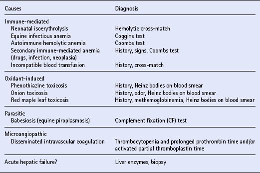
The hemolymphatic system

Chapter 9


The hemolymphatic system


Chapter contents


INTRODUCTION


Diseases of the hemolymphatic system are generally due to abnormal quantity and/or function of the normal cellular and humoral components of blood. In order to understand fully hemolymphatic diseases, a basic knowledge of normal blood production (hematopoiesis) and the hemostatic mechanism is necessary.


HEMATOPOIESIS


During postnatal life, hematopoiesis occurs in the bone marrow. Maturation is attended by recession of hematopoiesis from the shafts of the long bones and the replacement of red marrow by resting yellow marrow; however, active hematopoiesis continues throughout life in the epiphyses of long bones and in the flat bones of the skull, vertebrae, sternum, ribs and pelvis. Transition from yellow to red marrow can occur in response to increasing demand for erythrocytes via the glycoprotein hormone erythropoietin (q.v.).


The bone marrow is composed of differentiated blood cells and their recognizable precursors, undifferentiated progenitor cells, reticular cells, reticular fibers, endothelium-lined sinusoids and adipocytes. Hematopoiesis occurs in the intrasinusoidal spaces. All blood cells originate from a population of lymphoid-appearing cells, termed pluripotent stem cells, which give rise to committed progenitors of the lymphoid and myeloid series. Pluripotent stem cells (PPSC) are capable of slow self-renewal, whereas the committed progenitor cells differentiate into later stages and their absolute marrow numbers depend upon influx from the PPSC pool.


The amplification and differentiation of hematopoietic progenitors are regulated by polypeptide growth factors called colony-stimulating factors. Horse colony-stimulating factors have not been characterized but they are presumed to function similar to those described in humans.


Erythropoietin (Ep) is the most important erythropoietic growth factor. It is produced almost exclusively by the kidneys in response to tissue hypoxia. Committed erythroid stem cells become progressively more sensitive to Ep stimulation as they mature.


Under homeostatic conditions, marrow production of blood cells approximates the rate of cell destruction. The lifespans of equine erythrocytes and platelets are approximately 155 days and 7 days, respectively, and the circulating half-life of granulocytes is about 10.5 h. Increased use or peripheral loss of marrow-derived blood cells results in production amplification of the necessary component(s). Complete marrow failure initially causes enhanced susceptibility to infection due to the loss of granulocytes. Petechial hemorrhages and bleeding secondary to thrombocytopenia follow. Finally pallor and signs of anemia occur.


The lymphoid stem cell also arises from the PPSC, but lymphocyte precursor maturation and differentiation are considerably more complex than those of the other hematopoietic cells. Some of the developing lymphocytes are seeded into the thymus, where their maturation into T cells occurs. In mammals, B lymphocyte maturation proceeds in the bone marrow.


Although functionally distinct, T and B lymphocytes (q.v.) have a similar morphologic appearance. Circulating lymphocytes, most of which are T cells, are in transit to and from the secondary lymphoid organs where most lymphocyte interactions with antigenic challenge occur. The spleen constitutes the largest single mass of lymphocytes in the body. Lymphoid accumulations also occur in great abundance in lymph nodes and near mucosal surfaces.


The spleen functions only briefly in hematopoiesis during fetal life, but retains this potential into adult life. The unique architecture of the spleen and its fixed phagocytic cells bestow the important function of filtering from the blood aged or damaged cells, particulate debris and microorganisms. Hemoglobin from senescent erythrocytes is degraded and iron is stored within phagocytes until it is subsequently released into the plasma for reutilization during erythropoiesis. Splenic macrophages also perform a “pitting function” by removing inclusions such as Heinz bodies (denatured hemoglobin) or intra-erythrocyte parasites from erythrocytes.


In horses, the spleen serves as an important reservoir for erythrocytes. Adrenaline-responsive smooth muscle in the equine splenic capsule causes the packed cell volume (PCV) to increase during exercise or excitement by as much as 40%. Normalization of red blood cell (RBC) parameters after a period of excitement may require up to 1 h in physically fit horses. The spleen also serves as a dynamic reservoir of platelets such that, at any point in time, up to one third of the total blood platelets are retained in the spleen.


HEMOSTASIS


Hemostasis is a complex series of events that functions to arrest bleeding from damaged blood vessels and to maintain blood flow to all body tissues. The processes of coagulation and fibrinolysis, with their respective inhibitors, constitute hemostasis.


Coagulation


The cooperation of platelets and blood procoagulant proteins with the blood vessel wall provides the basis for coagulation, which culminates in the formation of fibrin. Immediately after injury, reflex vasoconstriction limits blood loss and the vessel wall provides a scaffold for fibrin clot formation. Endothelial cells release substances that activate or inhibit components of coagulation and fibrinolysis.


Platelets interact with a discontinuous vascular surface to form a plug that provides primary hemostasis. Platelets adhere to subendothelial collagen and then undergo activation, aggregation and the release reaction. An initial platelet release of adenosine 5′-diphosphate (ADP) into the extracellular environment in response to adhesion promotes primary aggregation.


Secondary irreversible aggregation is a consequence of thromboxane (TXA2) formation and secretion of platelet granule constituents. The phospholipid (PL) necessary at numerous steps during subsequent interaction of coagulant proteins is supplied by platelet factor 3. During and after platelet plug formation the platelet surface protects coagulant proteins from plasma anticoagulants and localizes clot formation. Platelets also prevent spontaneous hemorrhage into the skin and mucous membranes by maintaining “vascular integrity”.


Coagulation proteins (or coagulation factors) circulate in the peripheral blood as inactive enzymes (zymogens). The coagulation “cascade” is a self-amplifying series of proteolytic events in which a zymogen is transformed to a proteinase that effects the subsequent zymogen–proteinase transition. There are two mechanisms by which activated factor X is formed, then coagulation proceeds through thrombin and fibrin production via a single common path.


In the extrinsic pathway, factor Xa is formed via the action of tissue factor (TF), which accesses the circulation via inflammation or tissue necrosis. Fibroblasts in the blood vessel adventitia express surface TF constitutively, while vascular endothelial cells, tissue macrophages and blood monocytes are activated to produce TF by agonists such as endotoxin (q.v.).


The intrinsic pathway to factor Xa production is initiated when blood is exposed to a negatively charged surface such as the platelet plug and/or subendothelial collagen. Through association of the plasma protein factor XII (Hageman factor), prekallikrein and high molecular weight kininogen (HMWK), factor XIIa is formed which perpetuates the intrinsic cascade. Factor VIII generally circulates bound to von Willebrand factor (vWF) in an inactive form, but acquires procoagulant cofactor properties through limited proteolysis by thrombin or factor Xa.


Factor Xa forms a prothrombinase complex with factor Va, PL and Ca2+, which cleaves prothrombin to thrombin. Thrombin dissolves from the PL surface and cleaves fibrinogen to yield fibrin monomers, which spontaneously polymerize. Thrombin activates factor XIII, which crosslinks fibrin and increases the clot’s resistance to fibrinolysis.


Plasma anticoagulant proteins localize coagulation to the site of injury and thereby protect against generalized thromboses. One major anticoagulant mechanism involves antithrombin III (AT III), which accounts for approximately 75% of thrombin-inhibiting activity in plasma. AT III can also neutralize factors Xa, IXa, XIa and XIIa as well as kallikrein and plasmin. Heparin produces a conformational change in AT III that results in a 2000-fold acceleration of the latter’s inhibitory action.


The second major plasma anticoagulant mechanism is provided by protein C, a liver-derived vitamin K-dependent protein that circulates as a zymogen. When thrombin is complexed to endothelial cell thrombomodulin, it activates protein C rather than its other substrates. Activated protein C (APC) cleaves and inactivates factors Va and VIIIa in the presence of Ca2+ and PL. This action by APC requires another vitamin K-dependent cofactor, protein S.


Fibrinolysis


Fibrinolysis is activated simultaneously with coagulation and functions to prevent tissue ischemia by the continued presence of fibrin clots. The key fibrinolytic protein, plasmin, is formed from circulating plasminogen by the action of several plasminogen activators. The tissue plasminogen activator (tPA) is synthesized primarily by endothelial cells, and both tPA and plasminogen have a high avidity for fibrin. Stasis upstream from an occluded vessel is the primary stimulus for tPA release, and the additional uptake of tPA by the clot tips the balance of hemostasis toward fibrinolysis.


To ensure localization of plasmin activities to the fibrin clot, there is an efficient array of plasma antifibrinolytic proteins. Plasminogen activator inhibitor (PAI) rapidly binds and inactivates physiologic concentrations of tPA. Endothelial cells are the primary source of this PAI, although the release of PAI from platelets during coagulation may play an important role in prevention of premature clot lysis.


The inhibition of plasmin activity in the blood is instantaneously carried out by α2-antiplasmin (AP). During coagulation, AP competes with plasminogen for binding to fibrin, preventing spontaneous lysis of a normal clot.


ANEMIA


Anemia can be functionally defined as decreased oxygen-carrying capacity of the blood. Anemia occurs when the PCV is reduced below that which is considered normal for the horse’s age, breed and use. However, the PCV must be ≤0.30L/L before an individual can be classed as anemic.


Anemia develops due to one or more of three pathophysiologic mechanisms:


The bone marrow responds to blood loss and hemolysis by increased erythropoiesis (q.v.), thus the anemia is regenerative. A non-regenerative anemia ensues when the bone marrow does not replace senescent erythrocytes at a normal rate. A bone marrow examination (q.v.) is necessary to characterize accurately anemia in horses since peripheral signs of regeneration such as reticulocytosis and polychromasia rarely occur.


Clinical signs of anemia are due to reduced tissue oxygenation and include reduced exercise tolerance, depression, weakness, tachycardia, tachypnea and mucosal pallor. Signs are manifested at a higher RBC mass when anemia develops rapidly since a gradual onset of anemia allows physiologic compensation. A low-grade systolic murmur can sometimes be auscultated when the PCV drops below 0.15–0.18L/L. Fever, icterus and/or pigmenturia often accompany hemolysis. Epistaxis, hematuria or melena may signal chronic blood loss. Anorexia, lethargy and weight loss suggest an underlying disease process.


LABORATORY EVALUATION


The initial laboratory assessment of anemia includes a complete blood count (CBC), total plasma protein (TPP) and plasma fibrinogen. There are several unique features of the equine erythron that must be considered during evaluation of the CBC. A horse’s PCV cannot be accurately assessed during or after exercise, excitement or endotoxemia since these produce splenic contraction. Splenic contraction in response to acute hemorrhage (q.v.) also precludes evaluation of the severity of blood loss for the first 12–24 h. Small nuclear remnants called Howell–Jolly bodies are occasionally found in erythrocytes of normal horses and do not indicate increased erythropoiesis.


Characterization of anemia as non-regenerative in a horse is most accurately done by bone marrow examination. Aspirates to characterize marrow erythropoiesis are most easily obtained from the sternum using an 89 mm, 18G disposable spinal needle. Thin smears should be rapidly air dried, then stained with a modified Romanowsky (Wright) stain. The normal myeloid–erythroid ratio (M/E) ranges from 0.5 to 1.5 in horses, thus an M/E ≤0.5 classifies anemia as regenerative.


Equine erythrocytes are small (5–6 μm) and tend to adhere to each other to form a “stack” like coins (rouleaux). Marked rouleaux may be confused with autoagglutination, and this tendency for natural aggregation leads to rapid erythrocyte sedimentation from plasma. Equine plasma is normally quite yellow compared with that of other animals, due to the combined effects of blood carotenoids from green feed and the greater concentration of bilirubin. Fasting causes a marked increase in equine serum bilirubin and can result in clinical icterus (q.v.). There is no definitive explanation for fasting-induced hyperbilirubinemia in horses, but a net decrease in and/or competition for hepatic binding proteins (particularly ligandin) have been proposed. In consideration of hemolytic disease as a cause for icterus in horses, the influence of fasting must be determined.


Red cell morphology should always be evaluated as a routine part of the CBC. Precipitates of oxidized hemoglobin (Heinz bodies), parasites, or abnormal cell shape may aid in defining the cause of anemia. The RBC indices are not highly useful in horses since peripheral erythrocytes are nearly always mature. Hemolysis, in vivo or in vitro, causes an increased mean corpuscular hemoglobin (MCH) due to the presence of free plasma hemoglobin.


The leukogram and plasma fibrinogen are insensitive indicators of chronic inflammatory disease, which may be the cause of anemia. Chronic inflammation may be attended by a normal or only mildly elevated white blood cell (WBC) count in horses, while an intense erythropoietic response to anemia may result in neutrophilia. Both may cause a left shift.


Hyperfibrinogenemia is more indicative of the presence of inflammatory disease than the WBC count in up to 50% of affected horses. The TPP is useful in evaluation of hydration status, and may provide a clue to the cause for anemia. Reduced PCV in the presence of increased TPP, which may be due to hemoconcentration, suggests that the anemia is actually more severe than the PCV indicates. Mild anemia may be masked by dehydration in addition to splenic contraction. An increase in TPP due to hypergammaglobulinemia sometimes accompanies chronic infection. Reduction of both the TPP and the PCV is suggestive of chronic blood loss.


Generally, history, physical examination and baseline laboratory data allow categorization of the anemia as due to blood loss, hemolysis or inadequate erythropoiesis. Additional laboratory tests can then address the suspected cause for anemia. When more than one mechanism of anemia is suspected, a bone marrow examination (q.v.) is necessary to determine the presence or absence of erythroid regeneration.


BLOOD LOSS ANEMIA


The clinical and laboratory findings of blood loss are largely determined by whether this occurs acutely, chronically, externally or internally. Com-mon causes for acute blood loss in horses include trauma to the limbs, post-castration hemorrhage, rupture of a uterine artery at parturition or erosion of the carotid artery by guttural pouch mycosis (q.v.). Hypovolemic shock (q.v.), characterized by tachycardia, tachypnea, hypothermia, pale and dry mucous membranes, prolonged capillary refill time, cold extremities and muscle weakness, generally develops when blood volume is reduced by more than 30%. Compensatory mechanisms are triggered immediately in an attempt to maintain circulating blood volume.


The spleen masks the extent of blood loss for several hours post hemorrhage by injection of a concentrated mass of stored erythrocytes into the circulation. Catecholamines induce vasoconstriction and increase cardiac output. Plasma volume is expanded by fluid resorption and retention in the vascular system, which continues at a decreasing rate for up to 72 h. A decline in the TPP can be measured within 4–6 h of the insult, but a reduction of the PCV is usually not appreciated until 12–24 h post hemorrhage.


Diagnosis of acute blood loss is based on history of recent hemor-rhage, clinical signs and eventual development of anemia accompanied by hypoproteinemia. Hemothorax or hemoperitoneum (q.v.) must be documented by ultrasound examination or paracentesis and cytologic evidence of erythrophagocytosis


Treatment aims for acute blood loss are stopping the source of hemorrhage and maintaining circulatory blood volume. Rapid IV administration of large volumes (40–80 mL/kg) of sodium-containing isotonic crystalloid solution is necessary to control hypovolemic shock (q.v.). Preliminary data in horses suggest that smaller volumes of hypertonic saline (4–5 mL/kg 7.5% sodium chloride) may effectively reduce the pathophysiologic sequelae of experimental hemorrhagic shock; however, clinical studies are necessary to evaluate fully this therapy in horses. Hypertonic saline in the face of ongoing hemorrhage may be contraindicated.


Blood transfusion (q.v.) is indicated only when the erythrocyte mass is insufficient to maintain adequate tissue oxygenation. This occurs at a higher PCV if anemia develops rapidly. A PCV ≤0.20L/L indicates that all erythrocyte reserves have been depleted; however, blood transfusion is unnecessary if the PCV stabilizes at 0.12–0.15L/L. Relative renal hypoxia causes erythropoietin production that subsequently stimulates the bone marrow to begin replenishing erythrocytes within 4–6 days. The adult equine diet includes an excess of all necessary nutrients for erythropoiesis. Milk may contain insufficient iron. In general, iron-deficient states are extremely uncommon in horses and usually associated with chronic external blood loss. Anemia due to blood loss generally resolves within 4–12 wk.


Slow chronic blood loss allows the bone marrow to regenerate erythrocytes as they are lost. Anemia only develops once the rate of erythropoiesis is exceeded by the rate of hemorrhage. Gradual tissue hypoxia allows physiologic adaptation, thus clinical signs of anemia are generally masked until the PCV drops to ≤0.15L/L.


Causes of chronic gastrointestinal blood loss include: parasitism (particularly large strongylosis); gastric or duodenal ulcers; non-steroidal anti-inflammatory drug (NSAID) toxicosis; and neoplasia (particularly gastric squamous cell carcinoma) (q.v.). Melena is rare. Because chemical tests for fecal occult blood are not highly specific, diagnosis of chronic gastrointestinal blood loss should be supported by a high index of clinical suspicion and ruling out other sources of hemorrhage.


Blood loss from the upper respiratory tract is usually identified by epistaxis (q.v.). Guttural pouch mycosis, ethmoidal hematoma, fungal rhinitis and neoplasia (q.v.) are the most common causes for upper respiratory tract blood loss. Pulmonary bleeding subsequent to severe pneumonia, lung abscess or neoplasia is often occult and only recognized by finding hemosiderin-laden macrophages on cytology of a tracheal aspirate or bronchoalveolar lavage specimen (q.v.). Exercise-induced pulmonary hemorrhage does not result in anemia. Urogenital neoplasia or vascular anomalies (q.v.) may rarely induce chronic blood loss anemia via hematuria, which may be microscopic.


Blood loss anemia can develop due to hemostatic dysfunction. Severe coagulation factor deficiencies such as warfarin toxicosis, hemophilia A and other heritable coagulopathies (q.v.) generally induce clinically recognizable hemorrhage into joints or other body cavities. Acute hemorrhage may follow trauma or surgery. Thrombocytopenia and/or more complex coagulation disorders such as disseminated intravascular coagulation (DIC) (q.v.) may be associated with chronic anemia due to mucosal petechial and ecchymotic hemorrhages, epistaxis and occult blood loss from the bowel and urinary tract.


Treatment of chronic blood loss anemia includes identification and treatment of the primary disease process. Chronic external blood loss may lead to iron deficiency (q.v.) especially in foals, which have comparatively low body iron stores.


HEMOLYTIC ANEMIA


Hemolytic anemia is associated with erythrocyte destruction that exceeds the rate of normal bone marrow erythropoiesis. Intravascular hemolysis occurs in some disease processes, but hemolytic anemia is usually due to extravascular erythrocyte destruction and shortened intravascular lifespan.


Clinical manifestations of hemolysis vary with the rate of development and severity of anemia, as well as the underlying disease process. Clear hematologic evidence of anemia exists when icterus is caused by hemolysis. Acute intravascular hemolysis produces hemoglobinemia and hemoglobinuria, manifested as pink plasma and reddish-brown urine, respectively (q.v.). Constant or intermittent fever is not uncommon due to underlying infections or active erythrocyte destruction.


There are numerous causes and mechanisms for hemolytic anemia ( Table 9.1), which is regenerative anemia without hypoproteinemia. Intensified erythropoiesis is often associated with neutrophilia and regenerative left shift. Total and indirect bilirubin concentrations may be elevated. Other laboratory findings are determined by the cause of the anemia ( Table 9.1). In addition to a CBC, TPP and serum bilirubin, the diagnostic evaluation of suspected hemolytic anemia should include thorough blood smear examination, urinalysis, Coombs test and Coggins test (q.v.).


Treatment of hemolytic anemia is aimed at the primary disease process whenever possible. Massive hemolysis may warrant transfusion with blood, packed red cells or polymerized hemoglobin (q.v.).


IMMUNE-MEDIATED HEMOLYTIC ANEMIA


Antibodies bound to the surface of erythrocytes result in hemolysis. Erythrocytes coated with immunoglobulins or immune complexes are generally removed from the circulation by tissue-fixed macrophages in the spleen, liver and bone marrow (mononuclear phagocyte system, MPS). Complement-mediated intravascular hemolysis may occur if sensitizing antibodies are IgM or complement-activating IgG. Immune-mediated hemolysis may be primary (e.g. neonatal isoerythrolysis, transfusion reactions, q.v.) or secondary to infections, drugs or neoplasia.


Neonatal isoerythrolysis


Neonatal isoerythrolysis (NI) (q.v.) is a hemolytic syndrome in newborn foals mediated by maternal antibodies against foal erythrocytes (alloantibodies) absorbed from the colostrum. The disease most often affects foals of multiparous dams.


Etiology and pathology

In development of natural NI, the foal inherits from the sire and expresses an erythrocyte antigen (alloantigen) (q.v.) that is not possessed by the dam. Blood group incompatibility between the foal and dam is common but most alloantigens are weak immunogens under the conditions of exposure through parturition or placental leakage. However, factors Aa of the A system and Qa of the Q system are highly immunogenic, and antibodies to these induce approximately 90% cases of NI. Mares that are negative for Aa and/or Qa (approximately 19% and 17% of Thoroughbred and Standardbred mares, respectively) are at greatest risk of producing a foal with NI. There are reports of other inducing antigens, including Ab, Qb, Qrs, Qc, Db, Dq, Dc, Da, Ka, Pa and Ua.


Not all mares become sensitized to the incompatible alloantigen of their foals. This generally requires transplacental hemorrhage during a previous pregnancy with a foal possessing the same incompatible blood factor. An anamnestic response is usually necessary to induce a pathogenic quantity of alloantibodies. Ten per cent of Thoroughbred mares and 20% of Standardbred mares have “natural” antibodies to the Ca blood group antigen. Data suggest that these “natural” antibodies may suppress an immune response to other alloantigens since Aa-negative mares that have anti-Ca antibodies often do not produce antibodies to Aa of their foal’s erythrocytes if the latter also contain Ca antigen. Natural alloantibodies have not been associated with NI in horses.


Alloantibodies of sensitized mares are concentrated in the colostrum during the last month of gestation. The foal is normal at birth, since the mare’s complex epitheliochorial placentation does not allow in utero antibody transfer. Thus the final criterion for foal development of NI is ingestion of colostrum containing alloantibodies specific for foal alloantigens in the first 24 h of life. Ig-coated foal erythrocytes are rapidly removed from circulation by the MPS or lysed intravascularly via complement. Alloantibodies to Aa are potent hemolysins and generally cause a more severe clinical syndrome than other alloantibodies. Primiparous mares may be predisposed to NI by blood transfusion or other exposure to equine blood products.


Diagnosis

Lethargy, anemia and icterus during the first four days of life suggest the diagnosis of NI. Blood loss anemia is attended by pallor, and icterus due to sepsis or liver dysfunction is not associated with anemia. The definitive diagnosis of NI is based upon demonstration of alloantibodies in the dam’s serum or colostrum that are directed against foal erythrocytes. The hemolytic crossmatch between washed foal erythrocytes and mare serum with an exogenous source of absorbed complement (usually from rabbits) is the most reliable diagnostic test for NI. A number of qualified laboratories routinely perform this diagnostic service. Because some equine alloantibodies act only as hemolysins, agglutination tests may be falsely negative. The jaundiced foal agglutination (JFA) test between colostrum and EDTA-anticoagulated foal blood is a rapid screen for anti-red blood cell antibodies. False negatives may occur; however, if positive, this test may be used to determine when the colostrum is safe for the foal to nurse.


Treatment

The dam’s milk must be withheld during the first 24 h of life and the foal should be fed from an alternate milk source. A minimum volume of milk equivalent to 1% of the foal’s body weight should be fed every 2 h (e.g. a 50 kg foal should receive 500 mL of mare milk replacer q 2 h). The dam’s udder should be stripped regularly (<q 4 h) and the milk discarded. In most instances, NI is not apparent until the foal is >24 h of age when colostral antibodies have been depleted and/or the intestinal absorptive capacity for Ig has diminished. Withholding milk at this point is of minimal benefit.


Supportive care is paramount. Affected foals should not be stressed and exercise must be restricted to a box stall. IV fluids may be indicated to minimize the nephrotoxic effects of hemoglobin as well as to correct any fluid deficits and electrolyte/acid-base imbalances. Antimicrobials may be necessary to prevent secondary infections.


When a foal’s PCV drops to ≤0.12L/L, blood transfusion (q.v.) is warranted to prevent life-threatening cerebral hypoxia. Severe weakness, tachypnea and tachycardia indicate the need for transfusion, even at higher PCVs. Although erythrocytes from the dam are compatible with the foal, they must be washed free of plasma in order to prevent administration of additional harmful alloantibodies. Repeated centrifugation with replacement of plasma by saline is necessary to ensure clean packed RBCs. Since most field conditions do not allow safe utilization of dam erythrocytes, blood-typed individuals negative for Aa and Qa and free of alloantibodies are optimal donors.


The odds of finding a donor without Aa and/or Qa are higher in Quarter Horses, Morgans and Standardbreds than in Thoroughbreds and Arabians. If an untested donor must be used, a gelding with no prior history of blood transfusion is best. Two to four liters of blood or 1–2L of packed erythrocytes should be given over a 2–4 h period. These allogeneic cells have a very short lifespan and represent a burden to the neonatal MPS that may increase susceptibility to infection. In addition, these cells sensitize the foal to future transfusion reactions. Potential harm must be measured against the benefit in each particular situation.


Recently, bovine polymerized hemoglobin (Oxyglobin) has been used to save foals suffering from severe hypoxemia, prior to identification of an appropriate red cell donor. This ultrapurified hemoglobin improves oxygen-carrying capacity of the blood, but the half-life is likely to be <48 h. The recommended dose is 7.5 mL/kg (3 × 125 mL bags for a 50 kg foal).


Prognosis and prevention

The prognosis for NI in foals depends on the quantity and activity of absorbed antibodies and is indirectly proportional to the rate of onset of signs. Like most diseases, NI is much more effectively prevented than treated. Foals from any mare that previously produced a foal with NI should be provided with an alternate colostral source unless the sire has known blood type compatibility with the dam. Mares at risk of producing affected foals (negative for Aa and Qa alloantigens) may be identified by blood typing (q.v.). Stallions negative for Aa/Qa and suitable on the basis of other criteria may be difficult to identify. It is most reasonable to breed “at-risk” mares as desired then to screen their serum in the last month of pregnancy for the presence of alloantibodies. If alloantibodies (other than those to Ca) are detected, dam colostrum should be withheld and the foal provided with an alternative colostral source. The JFA test has been used to determine when it is safe for the foal to nurse.


Autoimmune hemolytic anemia


Autoimmune hemolytic anemia (AIHA) (q.v.) occurs when an individual forms antibodies that bind to its own erythrocytes. Primary AIHA is an idiopathic process wherein there is failure to recognize erythrocytes as self. AIHA may arise secondary to infections, drugs or neoplasia and is then referred to as immune-mediated hemolytic disease. Both types result in MPS destruction of erythrocytes with or without intravascular hemolysis. AIHA is uncommon in horses but can affect any age, sex or breed.


Clinical signs and laboratory findings

Affected horses have variably severe depression and/or exercise intolerance. Tachypnea and tachycardia are generally present and worsen with exercise. Fever depends on the severity of hemolysis as well as any concomitant disease. Mucous membranes may be moderately icteric. Pigmenturia is uncommon.


Rarely in AIHA, erythrocytes in a blood sample grossly agglutinate. Immune-mediated erythrocyte aggregation persists when the anticoagulated blood is diluted 1:2 in isotonic saline, whereas false autoagglutination due to severe inflammatory disease is easily dispersed. Since equine erythrocytes are small, spherocytosis is rare. Neutrophilic leukocytosis is common subsequent to bone marrow regeneration. Intravascular hemolysis causes pink plasma, increased MCH and pigmenturia. Indirect hyperbilirubinemia is common.


Etiology and pathogenesis

True autoimmunity results when B lymphocyte cell clones become abnormally reactive and fail to recognize “self”. Dysfunction of T suppressor cells or increased activity of helper T cells may have a role. Secondary AIHA is often caused by immune complexes that bind to erythrocyte membranes and mediate extravascular hemolysis. In other situations the erythrocyte membrane is altered by the primary disease process and is then no longer recognized as “self”. Finally, antigenic stimulation of the immune system may result in antibodies that cross-react with normal erythrocytes. Secondary AIHA has been described in horses with equine infectious anemia (EIA), Clostridium perfringens infection, injection site abscesses, lymphosarcoma, other internal neoplasia, protein-losing enteropathy, purpura hemorrhagica, and penicillin therapy (q.v.).


Diagnosis

Definitive diagnosis of AIHA is based upon demonstration of patient antibodies that react with their erythrocytes. Autoagglutination is diagnostic of AIHA provided false agglutination is excluded. If true autoagglutination is not evident, diagnosis is most accurately made by agglutination in a direct antiglobulin (Coombs) test (q.v.), performed by incubating washed patient erythrocytes with appropriate dilutions of antiserum to equine IgG, IgM and complement components. A false negative Coombs test may occur immediately following a hemolytic crisis, or when corticosteroid therapy has been initiated. The osmotic fragility test should not be used for definitive diagnosis of AIHA, since positive results occur during other diseases that compromise erythrocyte membrane function (e.g. oxidative insult).


Horses with AIHA should have a thorough diagnostic work-up in search of neoplasia and a Coggins test for EIA (q.v.).


Treatment

Any current medication should be immediately discontinued in an attempt to exclude the possibility of drug-associated AIHA. Necessary antimicrobials are replaced by the most chemically dissimilar substitute (e.g. do not replace penicillin with ampicillin). Horses with severe AIHA require corticosteroid therapy. All beneficial actions of corticosteroids are unknown, but they will reduce erythrocyte clearance by the MPS and impair autoantibody production. Dexamethasone (0.05–0.2 mg/kg, IV or IM once daily) seems to be most effective for initial therapy of equine AIHA. If the PCV does not stabilize within 24–48 h, the dose rate of dexamethasone should be increased to twice daily. The full effect of corticosteroid therapy often requires 4–7 days and blood transfusion may be necessary in the interim. Corticosteroids may worsen a primary infectious process and cause recrudescence of viremia in horses with chronic EIA.


Once the PCV stabilizes at >0.20L/L, the dose of dexamethasone can be decreased 10% every 24–48 h while carefully monitoring for relapse. When the dose of necessary dexamethasone is ≤0.04 mg/kg daily, therapy can safely be given orally. Corticosteroids should be tapered as soon as possible to the lowest necessary dose and given on alternate days for 1 wk before disconti-nuation. Dexamethasone can be discontinued when the PCV remains stable during therapy with 0.01 mg/kg q 24–48 h. Prednisolone or prednisone can be used in lieu of dexamethasone (approximately seven times greater dose given q 12 h); however, these are erratically absorbed after oral administration.


Horses that fail to respond to corticosteroids may be treated concomitantly with oral azathioprine at a dose of 2 mg/kg/day. Positive results are anecdotal. Blood transfusions are avoided unless absolutely necessary, since the transfused cells are often rapidly lysed by circulating antibodies. When anemia is life threatening, Oxyglobin therapy may be preferable (q.v.).


Equine infectious anemia


Equine infectious anemia (EIA) (q.v.) is a multisystemic retroviral disease of Equidae, characterized by immune-mediated hemolytic anemia. Horses of all types are affected, but the disease is most prevalent in the southeastern USA. Because of its importance, EIA will be briefly considered separately from other causes of AIHA.


Clinical signs and laboratory findings

Three forms of clinical disease have been described: acute, subacute to chronic, and chronic inapparent. Clinical signs of acute EIA occur 7–30 days after first exposure to the virus and include fever, depression, anorexia and mucosal petechial hemorrhages. Anemia is not seen at this stage. Horses that have been infected for more than 30 days show the more classic clinical signs of EIA, which include anemia, icterus, edema of the limbs and ventral abdomen, intermittent fever spikes and weight loss. Less common clinical signs are colic, ataxia, abortion and infertility. Deaths usually occur during this subacute to chronic form of the disease.


Most horses recover, but experience unpredictable periodic flare-ups of clinical disease. Severe environmental or management stresses and treatment with corticosteroids are known to induce recrudescence of EIA. A large number of EIA-infected horses do not show clinical signs. Although only detected by serology, these animals remain virus carriers and are a potential source of infection for other horses.


During acute EIA, thrombocytopenia is the first and most consistent laboratory finding. Leukopenia is often present with mild lymphocytosis and monocytosis. During the subacute to chronic stages of disease, the PCV and RBC count are reduced, with other laboratory indications of hemolysis. The Coombs test is often positive. Hypergammaglobulinemia, increased liver enzymes and proteinuria may develop. Chronic inapparent carriers and chronically infected horses between clinical flare-ups are hematologically normal.


Etiology and pathogenesis

The causative agent of EIA is a high molecular weight, non-oncogenic lentivirus of the retrovirus family. There is wide genetic variation of the antigenic properties of the viral envelope and there is drift, which interferes with development of protective immunity. Viremia is maintained persistently in infected horses, despite a detectable immune response.


The EIA virus is usually transmitted by blood from affected horses, although other body secretions could serve as source of infection. Natural transmission occurs through the interrupted feeding of horse flies (Tabanus spp.) and deer flies (Chrysops spp.). Horses showing clinical signs of EIA have a higher viremia and are much more likely to transmit disease than are inapparent carriers. In utero transmission of the EIA virus is possible.


The EIA virus multiplies in macrophages throughout the body and elab-orates viral proteins that stimulate humoral and cell-mediated immune responses. Acute disease is associated with massive virus replication and destruction of macrophages. The incubation period before clinical signs is usually 1–3 wk, but may be as long as 3 mo. A detectable serologic response is generally attained 2–6 wk after infection and positive serology persists indefinitely.


Signs of subacute-chronic EIA result from virus-induced immunologically mediated tissue damage. Immune complex attachment to erythrocytes via the viral hemagglutinin produces hemolysis. Resultant anemia is worsened by decreased bone marrow erythropoiesis. Periodic disease flare-ups are due to the immune response to viral antigenic drift.


< div class='tao-gold-member'>

Stay updated, free articles. Join our Telegram channel

Jul 8, 2016 | Posted by in EQUINE MEDICINE | Comments Off on The hemolymphatic system

Full access? Get Clinical Tree

Get Clinical Tree app for offline access