THE FOAL AT DELIVERY

Chapter 3


THE FOAL AT DELIVERY


EVENTS FOLLOWING DELIVERY IN THE NORMAL FOAL


It is important to recognize the normal events which take place so that the abnormal can be quickly recognized and appropriate corrective action taken. Unfortunately there are wide variations in the normal pattern of delivery and neonatal adaptation, which makes the decision to interfere difficult. The extent of veterinary intervention will often depend heavily on the experience of the owner, handler or groom. More experienced grooms may not need anything like as much support and will also often recognize problems earlier than the less experienced. Furthermore, the value of the foal and any complications during pregnancy will often dictate the role of the veterinary surgeon.


By virtue of the speed of natural delivery and the experience of most stud grooms, probably the majority of foalings attended by veterinarians are recognizably abnormal in some respect or another. The recognition of abnormalities may be more difficult than it appears, however. Intervention needs to be limited but also sensible – there is little point in leaving an obviously abnormal situation to sort itself out; conversely, interfering when there is no need to do so can do harm.


The foal is delivered during second stage labor. Normally one front foot is the first part of the foal to appear and this is closely followed by the other foot (somewhat behind the first) and then the foal’s nose and face. Usually the foal is delivered within the amnion, which most often breaks spontaneously as a result of opposing movements of the forelimbs and head. Failure of the amnion to tear at delivery is a common cause of asphyxia and an attendant must intervene to free the foal’s face and head. Foaling mares should not be interfered with without due cause because the time taken (normally around 20 minutes) for the foal to pass through the pelvis and being ‘squashed’ as it comes through helps to cause the final surge of cortisol required for final organ maturation in the foal.


The foal takes its first breaths with both chest and abdominal effort (usually within 30 seconds of delivery of the chest). There may be a series of initial gasps with neck arching. This is not a signal for intervention.


The normal respiratory pattern is rapidly established. The newborn foal will show fast deep breathing and this is accompanied by dramatic rise in blood oxygen (PaO2) which progressively increases with increasing muscular effort. There are significant other signs for a similar increase in respiration rate.


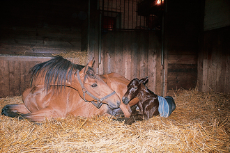
The mare will usually then undergo a period of tranquility (lasting up to 20–30 minutes) (Fig. 3.1) during which time the foal shakes its head and gains sternal recumbency with a ‘righting reflex’. Throughout this the mare remains quiet (usually in sternal recumbency) and will often vocalize to the foal.


This is not a sign for intervention unless the foal fails to become sternal within 5 minutes and/or has a low, labored respiratory pattern.


The foal may show strong blinking reflexes as hearing and vision are established. The foal may whinny on its own or in response to the mare. The foal’s head bobs up and down markedly and suckling responses with lips and mouth are present with increasing strength.


The foal then struggles and moves to the side of the mare. Usually the cord ruptures at this time, either from movement from foal or because the mare stands up. The cord usually ruptures about 6–8 minutes after delivery at a predetermined site (3–5 cm from the umbilicus). Shorter ruptures may have serious consequences including internal hemorrhage. Severe tension on the cord at the umbilicus may also cause serious internal bleeding (though this is very rare. There have been suggestions that premature rupture of the cord may deprive the foal of a significant volume of blood (25–30% of the foal’s blood volume can be lost1). However, studies measuring the haematocrit and haemoglobin in ‘premature cord rupture’ foals, and Doppler flow studies on blood flow within the umbilical vessels, have revealed that blood lost immediately after delivery was not clinically significant.2 Therefore, current opinion is that early (natural) rupture of the cord does not materially affect the adaptive period.


The mare nuzzles, licks and encourages the foal. In response the foal makes its first attempts to stand, usually within 30 minutes of delivery.


Normal foals will stand by around 45–90 minutes after delivery (often with apparent incoordination) and it may fall several times before establishing a steady stance and the ability to walk. A normal foal may take up to 2 hours before standing but the longer it takes the greater is the likelihood of a problem being present.


The foal then seeks for the mare’s udder; this is often aimless at first but with increasing accuracy. Once the teat is located a strong suckling reflex is stimulated. The first effective suckling usually takes place within 60–90 minutes of delivery. In response, the mare will ‘let down her milk’ and colostrum will be seen to stream from the teats.


Note: The significance of colostral ingestion cannot be overstated (see p. 14) and any foal that fails to find the teat and suckle within 2–4 hours (4 hours max) is abnormal and intervention is required.


After about 30–60 minutes (especially if a feed has been successfully obtained) the foal will lie down again. The foal may make energetic steps on rising again – it may jump up and down and may fall again. All foals have an inherent incoordination and may seem to be ataxic for the first 12–24 hours.


SUMMARY OF EVENTS TAKING PLACE DURING THE DELIVERY OF A FOAL


1. Recognition of correct time of interference – minimal interference is desirable.


2. Recognition of the amnion (as opposed to the chorioallantois or even rarely the bladder).


3. Cutting of the amnion is sometimes a desirable safety precaution: the foal’s nose can be uncovered and there is then less risk of asphyxia. However, it is important not to disturb the mare if at all possible while this is done.


4. The foal begins to breathe, taking deep and energy-demanding gasps, and a fast but regular breathing pattern is established. The foal will quickly achieve sternal recumbency with a lifted head; this position is conducive to good pulmonary inflation and function.


5. Cord rupture usually allows slight blood flow. Severe arterial blood loss from the umbilical end of ruptured cord requires immediate attention – clamp with sterile artery forceps or an umbilical clamp (plastic bag clamps are useful). The cord should not be tied with string, etc.


6. Navel treatment is given ideally as soon as the cord has ruptured or at least within 30 minutes of delivery. Recent work suggests that the best results are obtained by the use of chlorhexidine (0.5% solution) and that povidone iodine may not be as effective as was first thought.3 Ensure thorough soaking of the navel but avoid overhandling. Three dip treatments within the first 24 hours will probably be sufficient (see p. 76).


7. Nursing and ingestion of colostrum are essential within 2–4 hours (see p. 14). Intervention is needed if this is not achieved or if the quality of the colostrum is considered to be inadequate (see p. 14).


8. Full hygiene measures are imperative for anyone handling the foal – it is remarkable how few stud personnel have any concept of cleanliness when handling foals and parturient mares.


9. It is advisable to wear gloves and overalls which can be changed frequently on every occasion when dealing with neonatal foals (preferably protective clothing should be changed between different foals). Washing hands and changing overalls frequently also minimizes cross-contamination between mares foaling at the same time.


10. All reasonable hygiene precautions should be in effect at all times including the provision of freshly washed or disposable aprons/gowns for each mare/foal and for each stud. It is best to advise the stud to maintain a stock of these for their own personnel and for visiting vets.


SUMMARY OF NORMAL PARAMETERS AND POSTPARTUM CHANGES


PARTURITION INJURIES IN FOALS


Foals can sustain serious injuries during assisted and normal (unassisted) delivery. In the former cases injury is usually the result of overenergetic assistance or failure to correct the foal’s presentation and posture so that delivery can take place. In the latter cases the effects are harder to explain. A combination of the tendency to malpresentation and the high expulsive forces involved in natural delivery is capable of causing serious injury to both foal and indeed to the mare herself. The attending clinician (or stud manager) must try to ensure that the risks of injury are minimized because prevention is usually possible but treatment of the consequent injuries may be far less manageable.


As foals become more valuable there is an increasing tendency to panic during second stage labour and as mentioned above unnecessary interference can do more harm than good. However, in some cases assistance at foaling is required and the recent improvements in neonatal resuscitation may lead to the survival of foals that would otherwise have died. Therefore, a full understanding of injuries that can occur at parturition is important.


LIMB INJURIES


Fractures of the metacarpals (during forelimb traction) or metatarsals (during posterior presentations with hindlimb traction) can occur when obstetrical chains or ropes are used ill advisedly. Premature or dysmature foals may have poor calcification of the long bones (as well as the cuboidal bones of the knee/carpus and hock/tarsus) and even normal traction forces may then cause bone, joint or ligament damage. Such injuries are usually complicated by soft tissue trauma and may not be treatable.


Nerve trauma to the brachial plexus (most common, involving some degree of radial nerve paralysis) or lumbosacral plexus occurs most frequently during difficult deliveries. Signs include unilateral or bilateral hypotonia with depressed reflexes. Radial nerve paralysis presents as a foal unable to bear weight on the limb and unable to extend the carpus or digit. Treatment includes anti-inflammatory drugs (care of gastric ulcers) and splinting to allow the foal to bear weight and prevent limb contracture and additional trauma. The prognosis is dependent upon the location and severity of the trauma. Spinal root injury has a worse prognosis than plexus or peripheral nerve injury.


Prolonged traction can also cause serious vascular compromise that may only become obvious some days later. Skin damage (including bruising or even degloving injuries) can also occur.


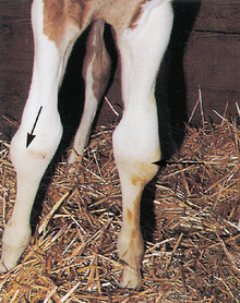
Rupture of the common digital extensor tendons (Fig. 3.2) may occur during forced extraction (they can also occur spontaneously; see p. 130).


Rupture of the caudal musculature of the hindlimbs (the gastrocnemius muscles and associated tendons) can occur from overzealous hindlimb traction, particularly if the hock is flexed while the stifle is extended. A foal with this condition will have a characteristic dropped hock (‘hamstrung’) posture, and a swelling is usually seen in the area of the gastrocnemius muscle. A complete disruption of the gastrocnemius will allow flexion of the hock when the stifle is in an extended position. Ultrasonography will identify muscle tearing. Treatment is extremely difficult and is unlikely in any case to result in restoration of athletic performance.


CRANIAL TRAUMA


Most cranial trauma is due to manipulations during parturition. Cranial fractures, jaw fractures and damage to eyes and mouth arise from careless or overenthusiastic attempts to deliver the foal. All handling involving the head and placement of snares must be performed very carefully using copious lubricant. There is never any need to exert extreme forces – if this is needed then an alternative approach must be sought.


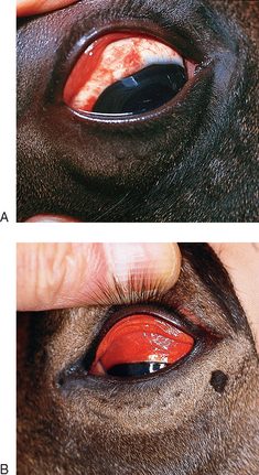
Some facial bruising is almost inevitable during handling and minor episcleral bruising or retinal hemorrhage (Fig. 3.3) is common even in normal foals; these do not usually require treatment. However, it is extremely wise to place such foals in a high risk category so that they are closely monitored for several days for signs of hypoxic ischemic encephalopathy (see p. 166) because the early signs can be misleading. It is particularly disappointing when extensive treatment (such as internal jaw fixation or skin sutures) is required to correct damage sustained during unsympathetic delivery attempts.


Jun 18, 2016 | Posted by in EQUINE MEDICINE | Comments Off on THE FOAL AT DELIVERY

Full access? Get Clinical Tree

Get Clinical Tree app for offline access