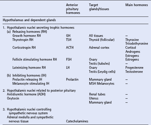
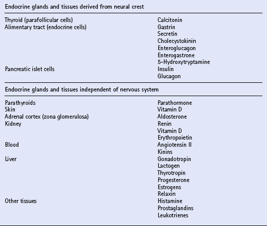
The endocrine system

Chapter 11


The endocrine system


Chapter contents


INTRODUCTION


The endocrine system comprises a complex and integrated system of glands and chemical substances (hormones) that function, along with the nervous system, to maintain homeostasis. Hormones have considerable influences on many of the metabolic processes of the body, and are involved not only in diseases that are commonly regarded as endocrine in nature, but also in such processes as growth and pregnancy, the response of the body to infection and trauma, and the development of neoplasms.


Much of the endocrine system is related developmentally, anatomically or functionally to the nervous system. The hypothalamus is part of the brain and is a most important part of the endocrine system. It controls directly the adenohypophysis (anterior pituitary gland), and indirectly (via the pituitary) several other glands, including the adrenal cortex, the gonads and the major part of the thyroid. Part of the hypothalamus extends into the neurohypophysis (posterior pituitary) to form a single anatomic and functional unit. Nuclei within the hypothalamus control the sympathetic nervous system, which includes the adrenal medulla.


Another group of endocrine tissues are derived embryologically from the neural crest, and are present as cells or clusters of cells within other organs; these include the parafollicular cells of the thyroid, the endocrine cells of the alimentary tract and the islet cells of the pancreas. Finally, there is a group of endocrine cells and tissues that is apparently independent of the nervous system; this includes the parathyroids, part of the adrenal cortex, and parts of other organs, tissues and blood. It is often said that the alimentary tract represents “the largest endocrine organ of the body”, and the complex endocrine and neural enteric systems have essential roles in the coordination of gut function, including motility, mucosal transport and local blood flow. The principal hormones and their sites of actions are summarized in Table 11.1.


Clinical endocrinology involves the study of how the body behaves in endocrine disorders and the application of the results to prevent, alleviate or cure endocrine disease. Unfortunately, the amounts of hormones usually present in the blood (especially the pituitary hormones) are extremely small, and the difficulty of measuring them has hindered the study of endocrine disorders. However, recent improvements in analytical techniques and in specificity and sensitivity of hormone assays have led, and continue to lead, to dramatic advances in the understanding of the normal and pathologic processes of endocrinology.


Diseases of the endocrine system often cause dysfunction of one or more of its parts. Hormones may be secreted in excess, causing syndromes of glandular hyperfunction, or in deficient amounts, causing syndromes of hypofunction. The overactivity or underactivity of one gland may adversely affect the function of another. Disorders may also arise because of a defect in the metabolism or breakdown of hormones, or the target tissues may become insensitive to the action of a hormone.


THE PITUITARY GLAND


INTRODUCTION


The equine pituitary gland measures approximately 2 cm × 1 cm and weighs from 1 to 3g. The terms anterior and posterior lobes are anatomically incorrect in horses, since the neurohypophysis (equivalent to the posterior lobe in primates) is embedded in and lies dorsal to the adenohypophysis (equivalent to the anterior lobe of primates). The adenohypophysis (80–85% of the total weight of the gland) is composed of endocrine cells derived from ectoderm. The neurohypophysis (15–20% of the total weight of the gland) consists of modified glial cells and axonal processes extending from nerve cell bodies in the supraoptic and paraventricular nuclei of the hypothalamus.


The adenohypophysis consists of three parts: the pars distalis, the pars intermedia and the pars tuberalis. Control of secretion from the pars distalis and pars tuberalis is by means of hypothalamus-derived releasing and inhibiting factors which are transported to the pituitary gland by local circulatory pathways. Control of secretion of the pars intermedia is by hypothalamus-derived neurotransmitters that are transported by axons directly into the pars intermedia.


The pars distalis is responsible for the secretion of numerous hormones, including growth hormone, prolactin, the gonadotropins luteinizing hormone (LH) and follicle stimulating hormone (FSH), pro-opiomelanocortin (POMC), adrenocorticotropic hormone (ACTH), corticotropin-like intermediate lobe peptide (CLIP), β-endorphin (β-END), thyrotropin (TSH) and melanocyte stimulating hormone (MSH). The pars intermedia is associated primarily with the synthesis of MSH and β-END.


The circadian rhythm of ACTH release arises mainly from corticotropin in the pars distalis. Increases in plasma ACTH lead to increases in circulating cortisol concentration. A diurnal pattern of ACTH and cortisol release occurs, with cortisol surges lasting 1–2 h starting between 02.00 and 04.00 h, resulting in peak plasma cortisol concentrations between 06.00 and 09.00 h. Circulating cortisol is largely bound to cortisol-binding globulin and has a half-life of 2–3 h.


The neurohypophysis comprises the pars nervosa, infundibular stalk and the pituicytes. Oxytocin and antidiuretic hormone (ADH) (also known as arginine vasopressin, AVP) are secreted by hypothalamic neurons arising in these nuclei, and are stored in the neurohypophysis.


The most common malfunction of the equine pituitary gland is associated with functional adenomas or adenomatous hyperplasia of the pars intermedia resulting in Cushing’s disease (q.v.).


DIABETES INSIPIDUS


Diabetes insipidus (q.v.) results from the decreased release of antidiuretic hormone (ADH) from the posterior pituitary (central diabetes insipidus) or lack of sensitivity of the kidney to actions of ADH (nephrogenic diabetes insipidus). The disease is most commonly associated with destruction of the posterior pituitary by compression from a tumor of the pars intermedia; however, rare cases of idiopathic diabetes insipidus have been recorded. The clinical signs include polydipsia and polyuria, with urine specific gravity <1.010 that fails to increase after a water deprivation test (q.v.). Serum ADH levels are low and remain unaltered by water deprivation.


Diagnosis may be achieved by using a water deprivation test and ADH response test. In the water deprivation test, access to all feed and water is denied and the bladder is emptied by catheterization. Urine specific gravity, body weight, blood urea and creatinine levels are monitored every 4 h for a maximum of 20 h. Water deprivation should be terminated if urine specific gravity rises >1.020, or if blood urea or creatinine levels increase above normal, or if there is clinical dehydration or a loss of more than 5% body weight. Horses in which urine specific gravity fails to exceed 1.020 during 20 h water deprivation are deficient in, or lack sensitivity to ADH. The ADH response test is performed by the IM injection of 40–60U pitressin (q.v.) following bladder evacuation and recording of urine specific gravity. Urine samples are collected 8, 12, 16 and 20 h post injection, and should exhibit maximal concentrations. Restoration of the ability to concentrate urine after ADH administration in an animal that failed to concentrate urine after water deprivation confirms central diabetes insipidus.


Treatment is by IM injections of desmopressin s.i.d. or b.i.d.; the dose must be adjusted to the individual requirements of the patient.


Failure to concentrate urine after water deprivation and ADH administration confirms nephrogenic diabetes insipidus, which may occur secondary to renal infection such as pyelonephritis (q.v.). This condition must be differentiated from the more common medullary washout that occurs in association with psychogenic water drinking. Animals affected by the latter condition are expected to show a normal urine concentrating response to water deprivation, although this may take the full 20 h or even slightly longer.


THE ADRENAL CORTEX


INTRODUCTION


The paired adrenal glands contain two anatomically and functionally distinct parts—the cortex and the medulla. The outer cortex, comprising 90% of the gland, surrounds the central medulla. The cortex is a zoned structure and is covered by a thin capsule, below which are isolated groups of glomerulosa cells. Most of the cortex is made up of the zona fasciculata and the zona reticularis, the latter adjoining the medulla.


The adrenal cortex produces three major types of steroid hormones: mineralocorticoids, glucocorticoids and androgens. The source of mineralocorticoids, aldosterone and in part deoxycorticosterone, is the glomerulosa cells. Aldosterone is involved in the regulation of sodium and potassium balance, and is under the control of the renin–angiotensin system (q.v.). Cortisol and the adrenal androgens (estrogens and progesterone) are derived from the fasciculata and reticularis cells. The only known important control mechanism is by means of ACTH. This adenohypophysis-derived hormone regulates adrenocortical growth; it also mediates the rate at which steroid biosynthesis occurs.


Cortisol and corticosterone are the principal glucocorticoids of horses. As with many other species, there is a circadian rhythm of adrenal activity, with peak secretion of glucocorticoids occurring in the morning. Levels of cortisol can be increased by stress (such as transport and surgery) and exercise. Normal term foals have a high plasma cortisol concentration and have a maximum response to exogenous ACTH during the first day of life. Premature foals have decreased adrenocortical function and the adrenal gland appears to be refractory to ACTH. Low cortisol levels have also been reported in chronic infections.


The most commonly recognized abnormality of the adrenal cortex in the adult horse is hyperadrenocorticalism (Cushing’s disease, q.v.) secondary to pituitary pars intermedia dysfunction. Hypoadrenocorticalism secondary to synthetic steroid administration also occurs, but there are few well-documented cases.


ADRENAL EXHAUSTION AND THE ACTH STIMULATION TEST


Adrenal insufficiency (also known as hypoadrenocorticalism or “steroid letdown syndrome”) is a poorly defined syndrome, sometimes associated with stress or poor performance syndrome (q.v.). Low circulating cortisol levels have been reported in endurance ride horses following a 100 mile (161km) ride, but abnormal responses to ACTH have not been determined and no deficiency in cortisol levels in racehorses affected by poor performance has ever been recorded. In fact, adrenal gland hypertrophy is more commonly observed at necropsy of racehorses than adrenal atrophy.


The adrenal glands are shock organs and can be damaged during endotoxemia, colic and anaphylaxis (q.v.). Long-term administration of exogenous corticosteroids or anabolic steroids can also lead to adrenal insufficiency if treatment is suddenly discontinued. The steroids suppress production of pro-opiomelanocortin (POMC) peptides by pituitary corticotropins, and the adrenal gland atrophies due to lack of ACTH stimulation. As little as 4 mg of dexamethasone may suppress the pituitary–adrenal axis for 18–24 h.


Clinical features of adrenal insufficiency include depression, anorexia, weight loss, poor haircoat and lameness. Serum biochemistry may be normal or show variable degrees of reduced levels of sodium and chloride, hyperkalemia and hypoglycemia.


Laboratory confirmation of adrenal insufficiency depends on an abnormal response to ACTH challenge; this may be performed using either ACTH gel or cosyntropin (synthetic ACTH). The test should be performed early in the morning. The protocols are summarized in Box 11.1.


Treatment of adrenal insufficiency includes rest and steroid supplementation.


CUSHING’S DISEASE


Introduction


Cushing’s disease (hyperadrenocorticalism) (q.v.) arises as a result of prolonged exposure to excess glucocorticoids, produced as a consequence of hyperplasia of the adrenal cortex. In the horse, this condition is almost exclusively associated with pituitary pars intermedia dysfunction, usually due to a functional tumor (adenoma).


The abnormal pituitary glands usually weigh 2–3 times the normal weight. The tumors cause varying degrees of compression of the pars distalis and occasionally infiltrate the neurohypophysis. Dorsal expansion of the tumor through the diaphragma sella can also lead to compression of the hypothalamus and optic chiasm, resulting in blindness and other neurologic deficits. The tumor cells have not been reported to metastasize.


Pars intermedia adenomas in horses contain markedly reduced amounts of dopamine (approximately 10% of normal), and it has been suggested that hyperplasia may arise as a result of loss of hypothalamic dopaminergic innervation. Currently, it is uncertain whether pituitary pars intermedia dysfunction in horses is the result of spontaneous pituitary disease (neoplasia) or the result of loss of dopaminergic innervation and thereby a primary hypothalamic disorder. If the latter mechanism is important, then there is justification for considering treating clinical cases with compounds with dopamine agonist action in order to limit the secretion of the active pituitary products.


Conventionally, and simplistically, it has been presumed that neoplastic pituitary glands secrete high levels of ACTH that result in hyperplasia of the adrenal cortices. However, the pathogenesis of equine Cushing’s disease is more complex, involving alterations in metabolism of other endocrine substances derived from the pituitary gland, including β-END, MSH and CLIP.


Although there are only limited data on the incidence of equine Cushing’s disease, it is recognized that the condition has a pronounced age distribution: essentially this is a disease of aged ponies and horses. The mean age of affected horses in a number of case series has ranged from 18 to 23 yr. The condition is very rare in horses <10 yr old, and the youngest recorded age is 7 yr.


The frequency of diagnosis of the condition has increased significantly over the past decade, but this is probably due to greater awareness and a larger population of older horses, rather than to an increasing prevalence. All breeds and types of equids (including donkeys) can be affected by Cushing’s disease, but ponies and Morgan horses may be at greatest risk. There is no apparent sex predisposition to the disease.


Clinical signs


Cases of equine Cushing’s disease may be presented for investigation of a fairly diverse range of complaints, including lethargy, dullness, weight loss, excessive thirst, chronic infections, abnormal haircoat, persistent sweating and chronic, refractory laminitis (q.v.). The classic clinical sign of Cushing’s disease is generally considered to be hirsutism (a long, curly haircoat that fails to shed). Despite the characteristic appearance of “classical” cushingoid ponies or horses, it is not uncommon for owners to have accepted many, or all, of these features as normal aging processes.


Most of the clinical signs of equine Cushing’s disease are explicable by the presence of increased amounts of plasma cortisol. For example, chronic, intractable infections are consequent upon prolonged immunosuppression, and laminitis (q.v.) arises from the effects of endogenous cortisol on the laminae of the foot. Commonly recognized infections include skin infections, recurrent subsolar abscesses, conjunctivitis, sinusitis, gingivitis and alveolar periostitis. Chronic insidious onset laminitis is seen in >50% of affected horses and is a common reason for euthanasia. The precise mechanisms by which hypercortisolism results in laminitis is poorly understood, but might relate to hyperinsulinemia and decreased glucose uptake by the laminar tissues.


High levels of plasma cortisol also stimulate protein catabolism, resulting in loss of body weight. Typically this is most pronounced in the epaxial and lumbar musculature such that affected animals appear dip-backed and/or pot-bellied. Despite the weight loss, the appetite of affected animals is usually normal, or may even be increased. However, dental abnormalities (q.v.) are common and can lead to painful mastication and quidding; this results in reduced feed intake that can also contribute to weight loss.


Other cortisol effects, seen less consistently, include oral ulceration and redistribution of fat. Deposition of fat occurs along the crest of the neck, over the tail head, and in the sheath of male horses. Another common site of fat deposition is above the eyes, producing prominent, bulging supraorbital fat pads.


Excessive thirst, or polydipsia, with increased urine output, is present in approximately one third of animals with Cushing’s disease. A number of different disease mechanisms may contribute to this polyuria–polydipsia (PU/PD) (q.v.). The enlarged pars intermedia may destroy the neurohypophysis by expansion resulting in partial neurogenic diabetes insipidus. Hypercortisolism may also result in central stimulation of thirst. In addition, PU/PD can arise through direct effects of cortisol on renal function causing increased glomerular filtration or antagonism of ADH at the collecting ducts. Cortisol also indirectly contributes to PU/PD via the osmotic diuresis effects of glucosuria, inevitable in hyperglycemic horses. The hyperglycemia of equine Cushing’s disease arises from the inhibition of the action of insulin to lower plasma glucose. Thus, PU/PD in equine Cushing’s disease may be a consequence of secondary diabetes insipidus (q.v.) and secondary diabetes mellitus (q.v.) within the same animal. Another possible urinary complication of Cushing’s disease is urinary tract infection, which may result in dysuria (q.v.).


Clinically, the most distinctive feature of cases of equine Cushing’s disease is the abnormal hair growth pattern which presents as an extremely long haircoat (hirsutism), frequently with a very curly appearance, and which is often retained for prolonged periods including into the summer months. During the first few years of the disease, the abnormal haircoat may be restricted to the lower jaw, base of neck and palmar/plantar aspects of the distal limbs. Later, generalized hirsutism may develop, and in some cases a dark haircoat may turn lighter in color. In some animals, the haircoat may be found to be matted and wet due to persistent sweating (hyperhidrosis) unrelated to exercise. The pathogenesis of hirsutism and hyperhidrosis (q.v.) is unclear but it has been suggested that it may be related to elevated POMC peptides or possibly due to the physical effect of the tumor causing pressure on the adjacent hypothalamus, thereby affecting the thermoregulatory center.


In a small proportion of cases, other clinical effects may arise from physical expansion of the neoplastic pituitary gland such as ataxia, seizures or blindness due to pressure on the optic nerves and/or their blood supply. Although interpretation of clinical findings such as lethargy or dullness must be somewhat subjective, it is not infrequent for these cases to appear abnormally docile, stoical or even somnolescent. It is possible that this changed behavior arises from raised levels of β-endorphins in plasma and cerebrospinal fluid (CSF).


Other clinical signs that have been reported in horses with Cushing’s disease include persistent lactation and infertility. These effects could be due to abnormal release of prolactin and gonadotropic hormones. Hypertrophic osteopathy has been reported in one pony with a pituitary adenoma. Breakdown of the suspensory apparatus and spontaneous fractures have also been described.


Diagnosis


In many instances, the clinical presentation and physical findings are virtually pathognomonic of equine Cushing’s disease. However, some cases may not manifest typical signs such that the diagnosis will require confirmation. The most consistent laboratory findings are hyperglycemia, glucosuria, raised plasma liver enzymes, mild anemia, absolute or relative neutrophilia and lymphopenia. Neutrophils may appear hypersegmented. On occasions, blood samples will appear lipemic due to raised levels of plasma triglycerides and cholesterol.


In principle, assessment of endocrine function in equine Cushing’s disease is appropriate in order to reach a definitive diagnosis, which may be particularly important prior to initiating treatment. In fact, the usefulness of endocrine function tests in horses is compromised to some extent by apparent wide variation in responses between individual animals, and a lack of studies that compare different tests or validate the results of tests with pathologic findings. The endocrine investigation is directed toward the assessment of function of the adrenal glands and the pituitary gland.


Adrenal cortical dysfunction may be assessed by the following:


Dexamethasone suppression test

The dexamethasone suppression test (DST) is considered to be the “gold standard” endocrinologic test for the diagnosis of equine Cushing’s disease by many workers. There is concern, however, that the administration of dexamethasone could exacerbate or induce laminitis (q.v.) although this complication appears to be extremely rare.


Several different protocols for the DST have been proposed, and two are summarized in Box 11.2. The overnight protocol is useful as a screening test, but the standard protocol allows more detailed assessment of the degree of loss of pituitary function.


In normal horses, the plasma cortisol will fall to 1μg/dL or less 19–24 h after the administration of dexamethasone. In affected horses, the cortisol levels show only a slight fall from basal levels.


Although the overnight DST is the most useful diagnostic test for Cushing’s disease in practice, both false positive and false negative results sometimes occur. Results always need to be interpreted along with clinical and other information.


Combined DST/ACTH stimulation test

The protocol for this test involves collection of a resting plasma sample and IM administration of 10 mg dexamethasone. A second plasma sample is collected 3 h later, followed by IV administration of 100IU of synthetic ACTH. A third blood sample is collected 2 h later.


The reason for performing the combined test is to suppress endogenous cortisol concentration in cushingoid horses to a value similar to normal horses before ACTH administration, and then to document an exaggerated response to exogenous ACTH. However, the combined DST/ACTH stimulation test is not recommended, since it does not allow evaluation of the rebound of cortisol seen in affected horses 24 h after dexamethasone administration. Also, adrenocortical hyperplasia is not a common feature of equine Cushing’s disease.

< div class='tao-gold-member'>

Stay updated, free articles. Join our Telegram channel

Jul 8, 2016 | Posted by in EQUINE MEDICINE | Comments Off on The endocrine system

Full access? Get Clinical Tree

Get Clinical Tree app for offline access