24 The control of pupil diameter
Abnormal pupil function in the pet can lead the owners to report large ‘black’ eyes, a staring expression, or sunken eyes which have ‘disappeared’ behind the prolapsed third eyelids and ptosis of Horner’s syndrome. The face may be described as having an ‘odd expression’. Dilation of conjunctival vessels from sympathetic paralysis is reported as a red eye. Anisocoria may be noticed.
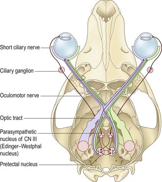
Pupils are normally symmetrically sized and shaped. Calm animals in an examination room have a pupil diameter of approximately 5 mm. Sympathetic drive actively dilates the pupils. Constriction is mediated by the parasympathetic branch of CN III via the ciliary nerves (Fig. 24.1). Structural change to the iris occurs with increasing age, and ocular pathology such as uveitis, glaucoma, lens luxation and synechia. Dyscoria describes a misshapen pupil. The feline iris is supplied by lateral and medial short ciliary nerves. If the lateral short ciliary nerve is affected, the pupil cannot constrict laterally and this gives a reverse-D-shaped pupil in the right eye and a D-shaped pupil in the left eye.