Tapeworms That Parasitize Domestic Animals and Humans
Learning Objectives
After studying this chapter, the reader should be able to do the following:
Key Terms
True tapeworm
Pseudotapeworm
Metacestode
Cysticercoid
Lappets
Egg packets
Oribatid grain mite
Cysticercus or bladderworm
Hexacanthr six toothed embryo
Coenurus
Unilocular hydatid cyst
Multilocular or alveolar hydatid cyst
Strobilocercus
Tetrathyridium
Operculated ovum
Procercoid
Plerocercoid
Sparganum
Hyatid cysts
Eucestoda (True Tapeworms)
Mice, Rats, Gerbils, and Hamsters
Intestinal Tract
Parasite: Hymenolepis nana and Hymenolepis diminuta
Host: Mice, rats, gerbils, hamsters, dogs, and humans
Location of Adult: Small intestine
Distribution: Worldwide
Derivation of Genus: Membrane covering
Intermediate Host: None necessary (H. nana): fleas, flour beetles and other arthropods (H. diminuta)
Transmission Route: Ingestion of infective fleas, grain beetle, or cockroach (H. diminuta); ingestion of infective egg or autoinfection (H. nana)
Common Name: Rodent tapeworm
 TECHNICIAN’S NOTE
While Hymenolepsis nana and Hymenolepsis diminuta are mainly parasites of mice, rats, gerbils, and hamsters, they have been found in dogs and humans.
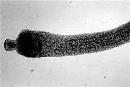
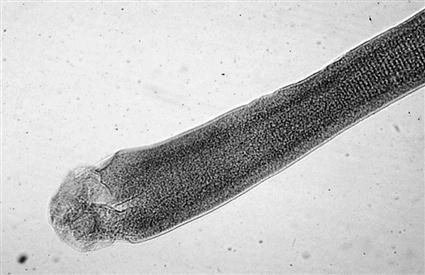
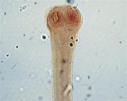
Hymenolepis nana and Hymenolepis diminuta parasitize mice, rats, gerbils, and hamsters. These tapeworms are small and slender. Adults of H. nana are 1 mm wide and 25 to 40 mm in length, and adults of H. diminuta are 3 to 4 mm wide and 20 to 60 mm in length. These true tapeworms reside in the small intestine of the rodent definitive host and are usually detected on postmortem examination of the small intestine. The scolex of H. nana has a ring of hooks on its anterior end; it has an armed rostellum (Figure 6-1). The scolex of H. diminuta has no hooks; it is unarmed (Figure 6-2).
The life cycle of H. nana is a direct life cycle, whereas H. diminuta requires an intermediate host for infection. The eggs of H. nana are passed in the feces and are swallowed by a host. The hexacanth enters the villus of the small intestine and matures into a nontailed cysticercoid. The cysticercoid returns to the lumen of the small intestine, attaches to the lining, and matures to adulthood. H. diminuta also passes its eggs in the feces, which are ingested by an intermediate arthropod host. The hexacanth embryo (embryo containing three pairs of hooks) matures into a tailed cysticercoid. The arthropod is ingested by a definitive host, and the cysticercoid attaches to the lining of the small intestine and matures into an adult.
 TECHNICIAN’S NOTE
Hymenolepsis nana is unique in that it is the only tapeworm that does not need an intermediate host for any developmental stage in its life cycle.
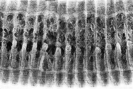
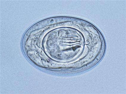
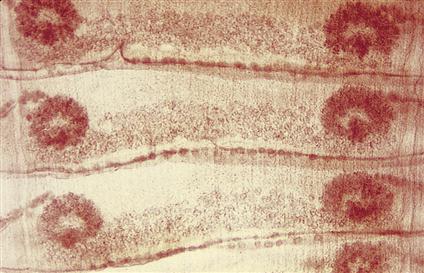
The eggs of Hymenolepis species may be detected on fecal flotation. Veterinary technicians should be aware that the eggs of this tapeworm are shed intermittently in the feces. Sometimes, individual proglottids may also be recovered, but these do not float. Figure 6-3 shows stained proglottids of H. diminuta. The oval egg of H. nana measures 44 to 62 µm × 30 to 55 µm (Figure 6-4). The egg of H. diminuta is more spherical and measures 62 to 88 µm × 30 to 55 µm. The embryo within the egg of both species measures 24 to 30 µm × 16 to 25 µm and contains three pairs of hooks (hexacanth embryo). Infected animals may be treated with niclosamide or praziquantel.
Both of these tapeworms have zoonotic potential. H. nana is unique in that it does not require an intermediate host; therefore it is directly infective to other rodents and to humans. Autoinfection by H. nana can occur when its eggs hatch in the small intestine of the host and subsequently infect that host. H. diminuta uses an insect (flea, grain beetle, or cockroach) as an intermediate host to complete its life cycle. The cysticercoid develops within the insect intermediate host (Figure 6-5).
 TECHNICIAN’S NOTE
Both Hymenolepsis nana and Hymenolepsis diminuta can infect humans.
Ruminants
Intestinal Tract
Moniezia species and Thysanosoma actinoides are true tapeworms that infect the intestinal tract of ruminants.
Parasite: Moniezia benedini and Moniezia expansa
Host: Cattle (M. benedini); cattle, sheep, and goats (M. expansa)
Location of Adult: Small intestine
Intermediate Host: Grain mites
Distribution: Worldwide
Derivation of Genus: Moniezia-to be single
Transmission Route: Ingestion of infective grain mite
Common Name: Ruminant tapeworms
Moniezia species.
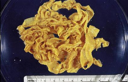
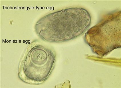
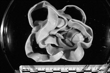
Moniezia species are long (up to 6 m) tapeworms found in the small intestine of cattle, sheep, and goats. Moniezia species are large tapeworms and can be up to 1.6 cm at the widest margins (Figure 6-6). The scolex of Moniezia is unarmed; it lacks an armed rostellum (Figure 6-7). Individual proglottids are very short and wide—“squatty.” Each proglottid contains two sets of laterally located genital organs and associated pores (Figure 6-8). These tapeworms produce eggs with a characteristic square or triangular shape. The eggs of both species possess a pyriform (pear-shaped) apparatus. Two species are common among ruminants: Moniezia benedini in cattle and Moniezia expansa in cattle, sheep, and goats. The eggs of both species can be easily differentiated using standard fecal flotation procedures. The eggs of M. expansa are triangular or pyramidal in shape and 56 to 67 µm in diameter. The eggs of M. benedini are square or cuboidal in shape and approximately 75 µm in diameter (Figure 6-9). The prepatent period for these tapeworms is approximately 40 days.
 TECHNICIAN’S NOTE
Moniezia species can be found with standard fecal flotation techniques. Moniezia benedini eggs have a square or cuboidal shape while Moniezia expansa eggs are triangular or pyramidal.
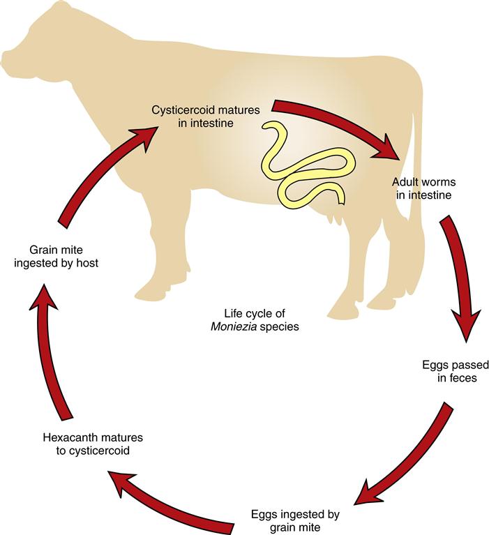
The metacestode, or larval, stage of Moniezia is the cysticercoid stage, which may be found within the intermediate hosts, oribatid grain mites. Intact proglottids and eggs are found in the feces of the ruminant definitive host. Mites become infected by ingesting the hexacanth embryo, which develops into the cysticercoid stage within the body of the grain mite. Ruminants become infected by ingesting cysticercoid-infected mites that infest the grain. The cysticercoid is a tiny, microscopic stage and probably will not be observed by the veterinarian (Figure 6-5). For every cysticercoid that is ingested by the ruminant, one adult tapeworm will develop in the small intestine of that ruminant (Figure 6-10). Large numbers of adults can cause rupture of the gut or obstruct the lumen of the intestines, especially in young animals. It is important that the veterinarian recognize the oribatid mite as the source of this tapeworm and understand the importance of effective tapeworm therapeusis (morantel, niclosamide, albendazole, fenbendazole, or oxfendazole) in cattle. Pasture rotation in addition to therapeusis is essential in greatly reducing the transmission of this parasite.
 TECHNICIAN’S NOTE
Pasture rotation is essential in reducing the transmission of ruminant tapeworms.
Parasite: Thysanosoma actinoides
Host: Sheep, goats, and cattle
Location of Adult: Lumen of the bile duct, pancreatic ducts, and small intestines
Intermediate Host: Unknown, proposed host is psocids insects
Distribution: North and South America
Derivation of Genus: Fringed body
Transmission Route: Ingestion of unknown intermediate host
Common Name: Fringed tapeworm of sheep and goats
Thysanosoma actinoides
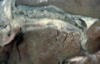
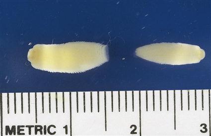
Thysanosoma actinoides is the fringed tapeworm found in the bile ducts (can cause bile duct obstruction), pancreatic ducts (can cause obstruction of pancreatic duct), and small intestine of ruminants (Figure 6-11). The adult tapeworm measures 8 mm × 15 to 30 cm and possess an unarmed scolex. As with Moniezia species, the proglottids are very short; however, these proglottids possess a unique feature: a very prominent fringe located on the posterior aspect of each proglottid. (Figure 6-12 shows the fringed adult T. actinoides.)
Eggs of this tapeworm occur in packets of 6 to 12 eggs, with individual eggs measuring 19 × 27 µm. These eggs do not possess a pyriform apparatus. The eggs can be found on standard fecal flotation procedures. Adults can be identified at necropsy.
 TECHNICIAN’S NOTE
These fringed tapeworms are unique among the adult tapeworms in that they possess a very prominent fringe along the posterior margin of each and every proglottid. The adults are also found in an unusual location for tapeworms, the lumen of the bile duct.
The metacestode (larval) stage of T. actinoides is the cysticercoid stage, which may be found within the proposed intermediate hosts, psocids. Psocids are primitive insects often associated with vegetation. These insects become infected by ingesting the hexacanth embryo, which develops into the cysticercoid stage within the body of the psocid. Ruminants may become infected by accidentally ingesting cysticercoid-infected psocids that infest vegetation. The cysticercoid is a microscopic stage and probably will not be observed by the veterinarian. For every cysticercoid that is ingested by the ruminant, one adult tapeworm will develop in the small intestine of that ruminant. It is important that the veterinarian recognize psocids as the source of this tapeworm and understand the importance of effective tapeworm therapeusis (niclosamide or praziquantel, as well as pasture rotation) in cattle.
Metacestode (Larval) Stages Found in Musculature of Food Animals
Parasite: Taenia saginata (Adult tapeworm)/Cysticescus bovis (metacestode [larval] stage)
Host: Humans
Location of Adult: Small intestine
Intermediate Host: Cattle
Distribution: Worldwide
Derivation of Genus: Flat band, bandage, or tape/bladder tail
Transmission Route: Ingestion of raw or undercooked infective beef
Common Name: Beef tapeworm of humans/beef measles, measly beef of cattle
Cattle may serve as intermediate hosts for a tapeworm of humans, Taenia saginata. The adult tapeworm is unusual among the Taenia species in that it does not have an armed rostellum like the rest of the species. Adults possess 14 to 32 lateral branches of the uterus within the gravid proglottid. The eggs are typical taeniid type ova with a striated embryophore (shell) surrounding an oncosphere with six hooklets inside. The adults can cause obstruction of the intestinal tract if present in sufficient numbers.
 TECHNICIAN’S NOTE
Taenia saginata is unusual in that it has an unarmed rotellum while all other Taenia species possess an armed rostellum.
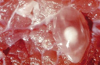
The larval stage for this tapeworm is a cysticercus, or bladder worm, called Cysticercus bovis. A cysticercus is a single invaginated scolex in a large, fluid-filled cyst, cavity, or vesicle. This condition in cattle is often referred to as “beef measles” or “measly beef.” These infective metacestodes are found in the musculature (skeletal and cardiac muscles) of cattle. If enough cysticerci are present in the muscle tissue, they can interfere with muscle function, produce pain, and myositis. Humans become infected with this zoonotic tapeworm by ingesting poorly cooked beef. (Figure 6-13 shows the cysticercus of C. bovis within beef muscle.)
Parasite: Taenia solium (adult tapeworm)/Cysticercus cellulosae (metacestode [larval] stage)
Host: Human
Location of Adult: Small intestine
Intermediate Host: Pigs
Distribution: Underdeveloped countries including Latin America, India, Africa, and the Far East
Derivation of Genus: Flat band, bandage or tape/bladder tail
Transmission Route: Ingestion of infective undercooked or raw pork
Common Name: Pork tapeworm of humans, measly pork, pork measles of swine
Pigs may serve as the intermediate host for a similar tapeworm of humans, Taenia solium. The adult possesses an armed rostellum with a double row of hooks. It is best identified by its 7 to 16 lateral branches of the uterus within each gravid proglottid. The eggs are typical taeniid type ova with a striated embryophore surrounding an oncosphere with six hooklets inside. In large numbers, the adult worms can cause intestinal obstruction. The ova can be found on standard fecal flotation. The adults can be identified by their characteristic lateral branches of the uterus.
 TECHNICIAN’S NOTE
The adult pork tapeworm of humans can cause intestinal obstruction in the human host if present in sufficient numbers.
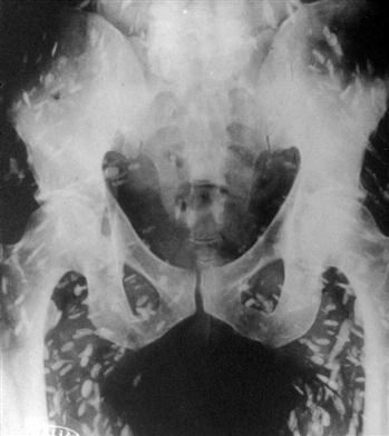
The larval stage for this tapeworm is a cysticercus, or bladder worm, known as Cysticercus cellulosae. This metacestode stage in pigs is often referred to as “pork measles” or “measly pork.” These metacestodes are found in the musculature (skeletal and cardiac muscles) of pigs. Humans become infected with this zoonotic tapeworm by ingesting poorly cooked pork. It is also important to note that if humans ingest the eggs of T. solium, the cysticercus can develop within their muscles (Figure 6-14) and within nervous tissue such as the brain, eye, and spinal cord.
The adult stages of the metacestode stages (C. bovis and C. cellulosae) are found in the small intestine of humans. Because humans may become infected by ingesting poorly cooked beef or pork, these are important zoonotic tapeworms.
 TECHNICIAN’S NOTE
Prevention of Taenia saginata and Taenia solium can be accomplished by thoroughly cooking the beef or pork to destroy the cysticercus within the meat, thus stopping the life cycle. Also, wash hands after handling raw beef or pork.
Metacestode (Larval) Stages Found in Abdominal Cavity of Food Animals
Parasite: Taenia hydatigena (adult tapeworm)/Cysticercus tenuicolis (metacestode [larval] stage)
Host: Dogs
Location of Adult: Small intestine
Intermediate Host: Cattle, sheep, goats
Distribution: Worldwide
Derivation of Genus: Flat band, bandage, or tape/bladder tail
Transmission Route: Ingestion of infective abdominal omentum of ruminants
Common Name: Canine taeniid/bovine bladderworm
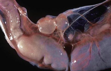
For Taenia hydatigena, an adult tapeworm found in the small intestine of dogs, the larval stage is a ping-pong-ball–sized, fluid-filled bladder called Cysticercus tenuicolis, which is usually attached to the greater omentum or other abdominal organs of the ruminant intermediate host (Figure 6-15) and is considered nonpathogenic to the intermediate host. The adult worm has an armed rostellum. The proglottids have a single, lateral genital pore. The eggs are typical taeniid-type ova with a striated embryopore surrounding an oncosphere with six hooklets inside. In large numbers, the adults can cause obstruction of the intestinal tract. To acquire this tapeworm, dogs become infected by ingesting the abdominal viscera of cysticercus-infected ruminants. For every cysticercus that is ingested by the dog, one adult tapeworm will develop in the small intestine of that dog. Diagnosis is made by finding the taeniid ova on standard fecal flotation. It is important that the veterinarian recognize the ruminant as the source of this tapeworm and understand the importance of preventing predation or ingestion of ruminant offal by the dog. Necropsy of the intermediate host will reveal the cysticercus stage in the abdominal cavity of the ruminant intermediate host.
 TECHNICIAN’S NOTE
Taeniid eggs can be found on standard fecal flotation.
Horses
Intestinal Tract
Parasite: Anoplocephala perfoliata, Anoplocephala magna, and Paranoplocephala mamillana
Host: Horses
Location of Adult: Small intestine, large intestine, and cecum (A. perfoliata); small intestine and occasionally stomach (A. magna and P. mamillana)
Intermediate Host: Grain mites
Distribution: Worldwide
Derivation of Genus: Unarmed head/Bearing an unarmed head
Transmission Route: Ingestion of infective grain mites
Common Name: Lappeted equine tapeworm (A. perfoliata) and equine tapeworm with large scolex (A. magna); Dwarf tapeworm (P. mamillana) collectively, equine tapeworms
 TECHNICIAN’S NOTE
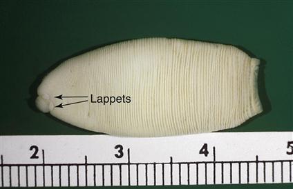
Anoplocephala perfoliata adults are easily identified by their morphologic features (lappets just posterior to the scolex) and the characteristic ovum with its pyriform apparatus.
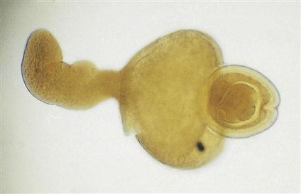
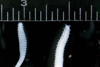
Anoplocephala perfoliata, Anoplocephala magna, and Paranoplocephala mamillana are the equine tapeworms. A. perfoliata is found in the small and large intestine and cecum; A. magna and P. mamillana are found in the small intestine and occasionally the stomach. A. perfoliata can measure from 5 to 8 cm in length and up to 1.2 cm in width. The scolex is oblong, 2 to 3 mm in diameter, unarmed, with very prominent lappets (two round protubences) behind each of the four suckers. The proglottids are wider than long, and each proglottid has only one set of male and female reproductive organs (Figure 6-16). A. magna can measure up to 80 cm in length and 2.5 cm in width. The scolex is large and oblong, 4 to 6 mm in diameter, unarmed but lacking the lappets of A. perfoliata (Figure 6-17). P. mamillana, also known as the dwarf tapeworm, is only 6 to 50 mm in length and 4 to 6 mm in width (Figure 6-18). The scolex is quite narrow. The adults of all three species can cause granulation tissue at the site of attachment in the intestinal wall.
The eggs of A. perfoliata are thick-walled, with one or more flattened sides measuring 65 to 80 µm in diameter; those of A. magna are similar but slightly smaller, measuring 50 to 60 µm. The eggs of P. mamillana are oval and thin-walled, measuring 51 × 37 µm. Eggs of all three species have a three-layered eggshell; the innermost lining is a pyriform apparatus. The hexacanth embryo can be visualized just inside the pyriform apparatus (Figure 6-19). Eggs of all equine tapeworms can be recovered using standard fecal flotation. The prepatent period for all three species ranges from 28 to 42 days.
 TECHNICIAN’S NOTE
All three species of equine tapeworms can be identified on standard fecal floatation by observing their triangular, three-layered-shell ova with the pyriform apparatus.
Stay updated, free articles. Join our Telegram channel

Full access? Get Clinical Tree


