Chapter 155 Surgical Correction of Patent Ductus Arteriosus
Ligation of the patent ductus arteriosus (PDA) in dogs and cats is a rewarding surgical procedure. When performed by an experienced surgeon, the combined operative/postoperative mortality rate is relatively low (8-10%) compared with surgery of other forms of congenital heart disease. The long-term prognosis after correction is excellent. However, like any thoracic or cardiovascular procedure, special attention to the details of anesthetic and surgical techniques is necessary for success. An accurate diagnosis is important, as is a thorough knowledge of the anatomy and physiology of the cardiovascular system.
ANATOMY AND PHYSIOLOGY
• The ductus arteriosus is a remnant of the left sixth aortic arch and connects the pulmonary artery and the descending aorta in the fetus and newborn. Its continued patency after birth results in left-to-right shunting of blood causing volume overload of the left atrium and ventricle. In severe cases, left ventricular failure may be present.
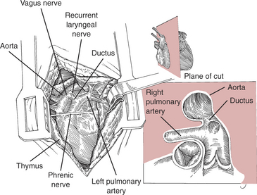
• The relationship of the descending aorta, main pulmonary artery, and right and left pulmonary arteries with the PDA must be appreciated (Fig. 155-1).
• The PDA may vary in size and shape but generally is approximately one-fifth to one-fourth the diameter of the aorta, or slightly smaller than the left main pulmonary artery. The PDA is usually short (0.5-1.0 cm), bridging the small distance between the aorta and pulmonary artery.
• The left vagus nerve lies over the ductus and is immediately underneath the visceral pleura. The recurrent laryngeal nerve arises from the vagus and courses caudal and medial to the PDA.
• Rarely, a persistent left cranial vena cava may be found coursing over the pulmonary artery. This does not pose a surgical problem in PDA ligation.
PREOPERATIVE AND PERIOPERATIVE CONSIDERATIONS
• Consult the chapter on congenital heart disease (see Chapter 154) for details of diagnosis and medical management.
• Institute conservative medical management before surgery when there is evidence of heart failure. More aggressive medical management may not benefit the patient as much as surgery.
• Mortality with surgery is higher when congestive heart failure (CHF), atrial fibrillation, or substantial myocardial failure is present. The mortality of non-surgically managed PDA patients also is high. Thus, if conservative therapy for heart failure is not effective in 24 to 48 hours, surgery combined with intensive medical management is the appropriate course of action in most patients.
• Administer IV fluids judiciously during anes-thesia and surgery, especially in animals with heart failure.
LIGATION OF THE PATENT DUCTUS ARTERIOSUS
Objectives
• Careful exposure and definition of the ductus arteriosus through a left fourth intercostal space thoracotomy. (The left fifth space may be indicated in cats.)


