CHAPTER 28 Surgical Correction of Abnormalities of the Female Reproductive Organs
SURGICAL CORRECTIONS FOR ABNORMAL PERINEAL CONFORMATION
Types of Abnormal Conformations
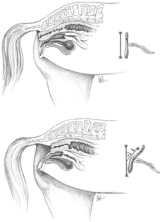
Vulvar Lips
Ideally, over 80% of the labia should lie in a vertical plane below the ischiadic arch of the pelvis. The Caslick index (La, where L = vulvar length and a = the angle of declination of the vulvar lips) has been developed to enable objective evaluation of the need for corrective surgery.1 Mares with an index above 100 may benefit from surgery and those with an index above 150 should definitely show improved fertility following surgery (Fig. 28-1).
Surgical Procedures
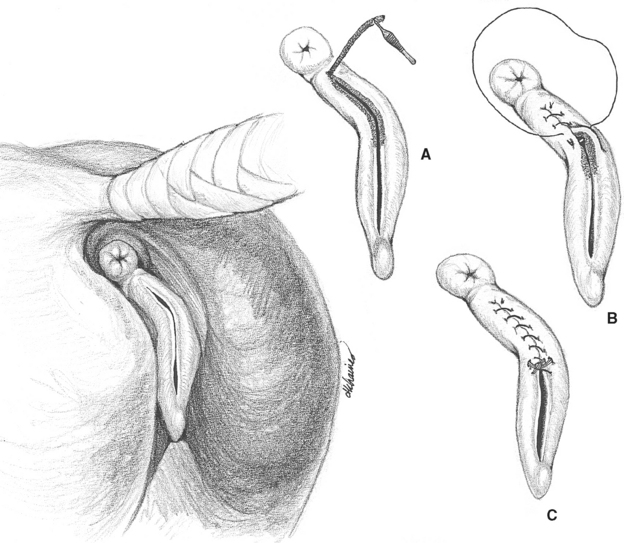
Episioplasty (Caslick’s Surgery)
Episioplasty2 is the surgical procedure used to reduce the length of the vulvar opening. Following is a summary of the steps involved:
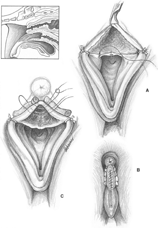
Perineoplasty
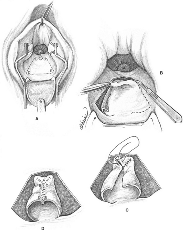
In mares that have a very loose vulvovaginal sphincter along with a cranially displaced vulva, episioplasty may not prevent pneumovagina. For these mares, perineoplasty may be performed by resecting a portion of the dorsal vaginal wall and apposing the denuded tissue, resulting in narrowing of the caudal vagina.3
SURGICAL CORRECTION OF URINE POOLING IN THE MARE
Surgical Procedures
Posterior Fixation of Transverse Fold
This technique is among the first described to extend the urethra.4 The urethral fold is pulled caudally, and its edges trimmed and sutured to incisions in the vaginal wall with No. 0 absorbable material. In most cases, caudal traction on the fold creates excessive tension and results in failure of the suture line. Additionally, it is difficult to achieve good apposition between the edges of freshened urethral fold and the vaginal wall (Fig. 28-4).
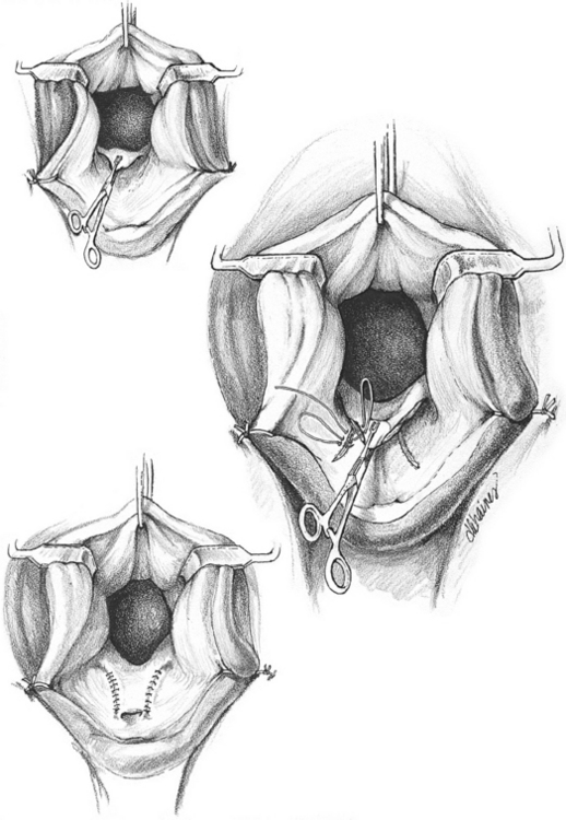
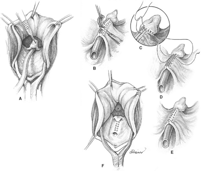
Urethral Extension (Shires Technique)5
After placement of a 30 French Foley catheter in the urethra (Fig. 28-5, A), interrupted horizontal mattress sutures (No. 0 synthetic absorbable material) are placed in the vaginal mucosa, sparing the catheter. As the sutures are tightened, folds of mucosa close over the catheter. Adequate mucosa must be present on the formed crest to allow mucosal tissue to be trimmed. Following excision, a simple continuous suture pattern is used to appose the cut edges. After completion of the procedure, the catheter is removed (Fig. 28-5, B to F).
Stay updated, free articles. Join our Telegram channel

Full access? Get Clinical Tree


