Chapter 91 Surgery of the Ovaries and Uterus
Surgical procedures performed on the uterus and ovaries include ovariohysterectomy, cesarean section, uterine biopsy, and rarely, ovariectomy. Uterine surgery usually is straightforward but requires sound basic surgery skills and a thorough understanding of the anatomy and physiology of the reproductive tract.
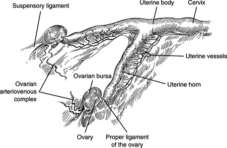
ANATOMY (Fig. 91-1)
Ovaries
• The suspensory ligament is the cranial continuation of the broad ligament and extends between the ventral third of the last two ribs and the ventral surface of the ovary.
• The proper ligament is a continuation of the suspensory ligament and extends from the caudal end of the ovary to the cranial end of the uterine horn.
• The ovarian arteriovenous complex (OAVC) lies on the medial side of the broad ligament and supplies the ovaries and the cranial portion of the uterine tube. The distal two-thirds of the OAVC is convoluted in the dog, similar to the pampiniform plexus in males.
• The left ovarian vein drains into the left renal vein; the right ovarian vein drains into the caudal vena cava.
Uterus
• The uterus consists of the cervix, body, and two uterine horns. Oviducts (uterine tubes) connect the uterine horns and ovaries.
• The uterus is attached to the dorsolateral wall of the abdominal cavity and the lateral wall of the pelvic cavity by paired double folds of peritoneum called broad ligaments.
• The round ligament is the caudal continuation of the proper ligament. The round ligament extends caudally and ventrally in the broad ligament and passes through the inguinal canal, terminating subcutaneously near the vulva.
OVARIOHYSTERECTOMY
Preoperative Considerations
• Elective sterilization is the most common indication for ovariohysterectomy. Ovariohysterectomy is the treatment of choice for most uterine diseases including pyometra, uterine torsion, cystic endome-trial hyperplasia, uterine rupture, and uterine neoplasia (see Chapter 90 for a description of these diseases).
• An alternative method of sterilization of a female dog or cat is ovariectomy without hysterectomy. Studies have shown no difference in long-term results, or complications, between ovariectomy and ovariohysterectomy. Ovariectomy may also be considered for the surgical treatment of small animal patients with a mass lesion involving the ovary, particularly if the owner wishes to maintain the reproductive status of the animal. However, if ovarian neoplasia is suspected, ovariohysterectomy coupled with a complete abdominal exploratory is indicated to ensure complete resection of the neoplasm and examination of all abdominal viscera for evidence of metastasis.
Surgical Procedure
Technique
4. Skin incision:
a. Dog: Make a ventral midline incision extending from the umbilicus to a point halfway between the umbilicus and the brim of the pubis.
b. Cat: Begin the ventral midline incision approximately 1 to 2 cm caudal to the umbilicus and extend the incision caudally 3 to 5 cm.
6. Locate the left uterine horn using the ovariohysterectomy hook or index finger. Displace the omentum and bowel cranially if necessary to find the uterus.
8. Grasp the ovary between the thumb and the middle fingers. Place the index finger as far proximal as possible on the suspensory ligament (Fig. 91-2A).
9. Place tension on the suspensory ligament by rotating the index finger caudally. Gradually increase tension on the suspensory ligament until the ligament stretches or ruptures.
10. Identify the OAVC. Using a Rochester-Carmalt hemostatic forceps (clamp), make an opening in the mesovarium immediately caudal to the OAVC in an area clear of vessels and fat (Fig. 91-2B).
11. Triple-clamp and transect the OAVC (Fig. 91-2C).
a. Double-clamp the OAVC with Rochester-Carmalt hemostatic forceps. Place the first clamp immediately proximal (toward the aorta) to the ovary and the second clamp approximately 5 mm proximal to the first. Place a third clamp across the proper ligament between the ovary and the uterine horn. Transect the OAVC between the middle clamp and the ovary (Fig. 91-2D).
12. Loosely place a circumferential ligature around the proximal clamp (Fig. 91-2F). Tighten the ligature as the clamp is removed. In this manner, the circumferential ligature is tightened in the groove of crushed tissue created by the clamp (Fig. 91-2G).
13. Place a transfixing ligature between the circumferential ligature and the transected end of the OAVC (Fig. 91-2H and I). A full ligature (circumferential) may be used instead of a transfixing ligature in young cats or small dogs.
14. Grasp the OAVC distal to the ligature (without grasping the ligature) with thumb forceps, remove the middle clamp, and inspect the OAVC for bleeding. If bleeding occurs, place a second circumferential ligature on the OAVC proximal to the first.
15. Follow the left uterine horn distally to the bifurcation, locate the right uterine horn, and follow the right uterine horn proximally to the right OAVC.
17. Transect the broad ligament.
a. In most preparturient animals, the broad ligament can be manually separated. Make an opening in the broad ligament adjacent to the uterine artery and vein close to the cervix (Fig. 91-2J). Place four fingers through the opening in the broad ligament and grasp the entire broad ligament, including the round ligament (Fig. 91-2K). Pull the broad ligament cranially (not ventrally) until the broad ligament and the round ligament are free (Fig. 91-2L).
19. Divide the uterine body after two ligatures are placed (Fig. 91-2M through O). Remove the entire uterus proximal to the cervix.
20. Evaluate the OAVC pedicles and the uterine body for bleeding prior to abdominal closure. The left and right OAVC are located immediately caudal to the caudal pole of each respective kidney.
a. Locate the left OAVC pedicle by retracting the descending colon medially, exposing the left paralumbar gutter.
b. Locate the right OAVC pedicle by retracting the duodenum medially, exposing the right paralumbar gutter.
Postoperative Care
• Dogs with pyometra frequently have renal dysfunction without associated morphologic abnormalities, and they may be azotemic, oliguric, or anuric. Monitor renal function and maintain hydration after surgery. Diuresis with crystalloid fluids administered intravenously for at least 24 to 36 hours after surgery is advisable. Placement of a urinary catheter will assist with monitoring urine output (see Chapter 90 for more information on pyometra).
Stay updated, free articles. Join our Telegram channel

Full access? Get Clinical Tree


