Chapter 72 Surgery of the Liver and Biliary Tract
Surgery of the liver and biliary tract is commonly performed in small animals and can be very challenging. Animals frequently are presented with surgical diseases of these organs, such as liver tumors and obstruction or infection of the biliary system. Some of the procedures described in this chapter require specialized training and facilities. Others, such as liver biopsy, partial hepatectomy, and simple exploration of the biliary tract, can be performed in a standard veterinary practice. Regardless of the procedure, preparation for the surgery by reviewing anatomy, pathophysiology, and specific techniques is very important for a successful outcome. Preparation of the patient prior to surgery also is extremely important because most of these diseases have serious metabolic effects.
SURGERY OF THE LIVER
Anatomy
Liver Lobes
Liver Attachments
Blood Supply
Portal Vein
Hepatic Arteries and Veins
Preoperative Considerations
Liver diseases cause a variety of significant hematologic and metabolic disorders in animals. Prior to surgery, perform appropriate diagnostics to confirm the disease and check for involvement of other organs. (See Chapter 71 for diagnosis of liver problems.)
Of particular concern to the surgeon are the following potential problems:
Diffuse hepatic disease may cause serious metabolic problems that can be compounded by anesthesia and surgery. Analyze liver function tests, such as serum bile acid and blood ammonia concentrations (see Chapter 71), to determine the animal’s ability to undergo anesthesia and surgery.
Liver Biopsy and Partial Hepatectomy—Surgical Procedure
Equipment
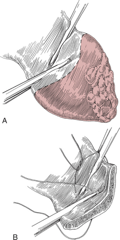
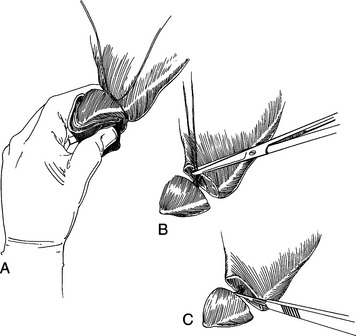
Technique

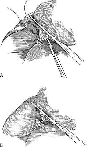
Figure 72-2 Partial hepatectomy. Lesions at the periphery of the liver lobe can be removed by placing crushing clamps across the lobe, proximal to the lesion, and then cutting distal to the clamps. Use absorbable sutures to ligate vessels proximal to the clamps. Tie these ligatures tightly as in the guillotine method for liver biopsy (see Fig. 72-1).
Postoperative Care and Complications
SURGERY FOR PORTOSYSTEMIC SHUNTS
Portosystemic shunts are abnormal vascular communications between the portal vein and a systemic vein. Diagnosis and medical treatment of this condition are discussed in Chapter 71. Animals with portosystemic shunts can initially improve with medical management. However, the definitive and most effective therapy for long-term resolution of clinical signs is surgical attenuation of the shunt vessel. With recent advances in the intraoperative and postoperative management of these animals, morbidity and mortality rates associated with surgery have decreased to very acceptable levels.
Anatomy
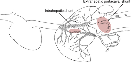
Portosystemic shunts are divided anatomically into extrahepatic and intrahepatic (Fig. 72-4). Definition of shunt anatomy preoperatively is important because intrahepatic shunts are much more difficult to correct surgically than are extrahepatic shunts. Also, familiarity with the anatomy helps decrease surgical time and patient morbidity.
Stay updated, free articles. Join our Telegram channel

Full access? Get Clinical Tree


