RESTRAINT/POSITION
Standing sedation or anesthesia may be used.
TECHNICAL DESCRIPTION OF PROCEDURE/METHOD
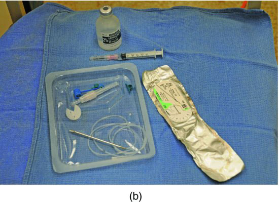
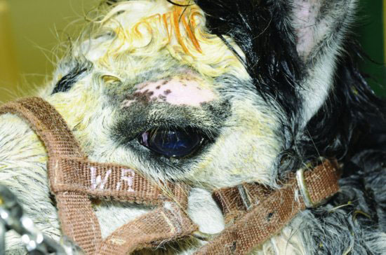
The animal must be sedated or anesthetized. The upper eyelid must be clipped from just above the eyelashes to 1 inch above the orbital rim (Figure 71.2). Surgical prep of this skin is done using alternating iodine scrub and sterile saline wipes (alcohol should be avoided) for a minimum of three repetitions. A local block should be placed at the orbital rim and another at approximately 1 cm cranial to the orbital rim using up to 1 mL of bupivacaine or 2% lidocaine (Figure 71.3). Apply a small amount of proparacaine to the cornea for topical anesthesia (Figure 71.4). The trocar needle of the lavage system is loaded with the nondisc end of the lavage tubing. The rest of the tubing is held in the gloved hand. The trocar needle is held along the index or middle finger of the dominant hand. The index or middle finger of the dominant hand is slid up the inside of the upper eyelid with the needle facing bevel out. At the fornix of the upper eyelid, as far proximally as possible the trocar needle is pushed through the upper eyelid at the orbital rim being careful to keep the tubing in the needle (Figure 71.5). Needle and tubing are advanced and pulled completely through the eyelid being careful to protect the cornea from needle trauma with the gloved finger. When the needle has been passed completely through the eyelid, the needle is removed from the tubing and the tubing is advanced through the eyelid until the disc at the end of the tubing is pulled firmly into the upper eyelid fornix, again, being careful to protect the cornea from damage associated with the plastic disc, using the gloved finger (Figure 71.6). When the disc is in place, the tubing is dried, and either the provided plastic rings are used or white tape wings are used to secure the tubing to the upper eyelid. This author’s preference is to use white tape to create wings that can be sutured to the skin (Figure 71.7), but the kit comes with a plastic loop that can be used. This plastic securing device must be used correctly; the tubing is passed through the small holes over the top of the large loop, and the suture is placed through the large loop to the head. Additional stay sutures can be placed if security is questionable, at the veterinarian’s discretion. The kit provides a catheter and an end cap to be placed in the distal end of the tubing (which can be cut shorter if desired). The end cap must be placed onto the catheter before placing the catheter into the tubing. Care must be used to avoid puncturing the tubing with the introducer needle, pulling the sharp portion into the catheter, after it has been introduced to the tubing is helpful. The catheter is advanced all the way to the hub, the needle is discarded, the end is capped, and the remaining tubing is secured to the animal (Figure 71.8).
Stay updated, free articles. Join our Telegram channel

Full access? Get Clinical Tree


