CHAPTER 29 Staphylococcal Infections
ETIOLOGY
The genus Staphylococcus consists of a group of approximately 50 different species and subspecies of gram-positive cocci, many of which are opportunistic pathogens in horses. Staphylococcal species are typically divided based on the ability to produce coagulase (Box 29-1). They are also often divided into major and minor pathogenic species based on the incidence and nature of disease. Most major pathogenic species are coagulase positive, and the two most important staphylococci in equine medicine are the coagulase-positive species Staphylococcus aureus and S. intermedius.1 Coagulase-negative staphylococci (CoNS) can be pathogenic, but infection with these species is most common in hospitalized or otherwise compromised hosts.
Box 29-1 Examples of Staphylococcus Species Based on Coagulase Production
| Coagulase positive | Coagulase negative |
|---|---|
Many species are part of the normal microflora, particularly of the nasal passages and skin. In humans, S. aureus is most often found in the anterior nares, and studies have reported colonization rates of 29% to 38% in healthy individuals.2,3 The majority of people intermittently carry S. aureus in their nasal passages, whereas some may never be colonized and others are persistently colonized. Staphylococcus epidermidis is the predominant staphylococcal species on skin surfaces, and other CoNS can be found on a variety of body surfaces. Staphylococcus intermedius is uncommon in humans.
Body site colonization has not been evaluated as extensively in horses. Nagase et al.4 reported isolation of staphylococci from the skin of 89.5% of horses. Staphylococcus sciuri was the predominant species, with lesser numbers of S. xylosus, S. saprophyticus, S. hominis, S. epidermidis, and S. capitis. Staphylococcus sciuri and S. xylosus predominated in another study of normal skin.1 Other studies have reported the predominance of CoNS or nonhemolytic staphylococci on the skin over joints.5,6 Fewer studies have evaluated staphylococcal colonization of other body sites. Yasuda et al.7 reported isolation of S. sciuri or S. lentus from the nares and pasterns of 13 of 100 mares in Japan. Staphylococci, predominantly S. aureus and CoNS, may be found as part of the conjunctival microflora of normal horses.8,9
The most significant multidrug-resistant Staphylococcus species in human medicine is methicillin-resistant S. aureus (MRSA). MRSA is one of the most important nosocomial pathogens and is associated with increased morbidity, mortality, duration of hospitalization, and treatment costs.10,11 Mortality rates of 50% for bloodstream infections and 33% for MRSA pneumonia have been reported, and outbreaks in medical facilities are a concern.12 Methicillin resistance in MRSA is mediated by production of an altered penicillin-binding protein (PBP) that possesses low affinity for beta (β)-lactam antimicrobials. Clinically, MRSA isolates are resistant to all β-lactam antimicrobials and are frequently resistant to a variety of other antimicrobial classes, and few treatment options may be available.
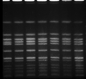
Recently, it has become evident that MRSA is an emerging pathogen in horses (Fig. 29-1). Hartmann et al.13 reported postoperative MRSA infection in a horse, and Seguin et al.14 reported nosocomial MRSA wound infections in 11 horses over a 13-month period at a teaching hospital. Indistinguishable MRSA isolates were also taken from three hospital personnel in the latter study, and it was speculated that hospital personnel were the source of equine infection. Weese et al.15 reported MRSA colonization or infection in 79 horses in a teaching hospital and on horse farms. A later study identified MRSA colonization in 46 of 972 (4.7%) horses on farms in Ontario and New York.16 As part of an equine MRSA screening program, community-associated MRSA colonization was identified in approximately 2.5% of horses admitted to a teaching hospital, and colonization at the time of admission was a risk factor for development of MRSA infection during hospitalization.17
Methicillin resistance is common among coagulase-negative Staphylococcus spp., including commensal species, although methicillin-resistant CoNS are not considered to be as important clinically as methicillin-resistant coagulase-positive staphylococci. Methicillin-resistant CoNS were isolated from the nares or skin of 29.5% of healthy horses in one study, with S. epidermidis being the most common species.18
PATHOGENESIS
Staphylococci can possess an impressive array of virulence factors that enable them to colonize and cause disease. Most virulence factors have been identified in S. aureus, but some may also be found in other staphylococci. Exozymes, including coagulase, protease, hemolysins, hyaluronidase, collagenase, lipase, and nuclease, facilitate colonization and growth in vivo and are possessed by virtually all S. aureus isolates and variably by other species.19 Additional virulence factors may be possessed, particularly by S. aureus. Staphylococcal enterotoxins (SEs), also classified as “pyrogenic staphylococcal superantigens,” are produced by various coagulase-positive staphylococci and are primarily associated with food poisoning in humans.19,20 These toxins are typically associated with S. aureus; however, SE genes have also been found in S. intermedius. SEs have been identified in S. aureus and S. intermedius of equine origin,21–23 but the role of SE in equine disease is unclear.
Toxic shock syndrome toxin (TSST-1) is another pyrogenic staphylococcal superantigen and is associated with staphylococcal toxic shock syndrome in humans.19 TSST-1 is unique in its ability to cross intact mucosal surfaces, and systemic disease can result from localized infections. Toxic shock syndrome associated with TSST-1 has been reported in a horse.24 Exfoliative toxin (sET) is responsible for skin exfoliation in staphylococcal scalded-skin syndrome in humans25 and has been associated with skin infection in a horse.26 The Panton-Valentine leukocidin (PVL) is associated with tissue necrosis and is emerging as an important virulence factor, particularly in community-associated MRSA infection.27–29 This toxin has not yet been identified in any tested equine isolates.15
The mere presence of staphylococci in or on a host does not mean that disease is occurring or will occur. Staphylococci are opportunists, and one or more risk factors typically must be present before disease can occur. Risk factors for development of clinical infection in humans include surgery, trauma, concurrent infection, skin lesions, and immunocompromise.27,30,31 Other risk factors presumably exist and may not be obvious. This is highlighted by the increasing incidence of community-associated MRSA infection in people without traditionally accepted risk factors.32–34 Risk factors for development of staphylococcal disease in horses have not been adequately evaluated but are likely similar to those reported in humans.
The source of staphylococci in equine infection is often not identified. Potentially pathogenic staphylococci may originate from the endogenous microflora of the infected horse, in-contact horses, or in-contact humans. The relative importance of these potential sources is unclear. Endogenous MRSA is a source of infection in some horses,17 but whether this is true for other staphylococci is unclear. Transmission of staphylococci between horses is probably common based on the behavior of the species, which involves frequent contact with the nares of other horses and exploratory behavior that results in contact with feces or items (e.g., buckets) that have been in recent contact with other horses. The clinical significance of this is unclear, although transmission of MRSA between horses with subsequent development of disease has been identified.15
CLINICAL FINDINGS
The clinical presentation of staphylococcal infection can be quite variable and depends on the location of infection, virulence of the involved strain, and host factors. Severity of disease can range from mild local infection to septicemia and toxemia. Many studies have reported only a general classification of “staphylococci,” hampering the ability to identify the relative importance of varying species. In general, most staphylococcal infections are characteristic opportunistic infections, particularly involving wounds and surgical sites, although primary infections of a variety of body sites have been reported. Staphylococci are important causes of septic arthritis after intraarticular injection, with one study isolating staphylococci from 86% of cases.35 It is unclear whether origin of these infections is inadequately prepared skin or the hands of veterinary personnel.
Coagulase-Positive Staphylococci
Staphylococcus aureus is believed to be the most important Staphylococcus species in equine medicine, and most information regarding equine staphylococcal infections involves this organism. Infections of wounds and incisions, subcutaneous tissues (cellulitis), bones and joints, and skin are most frequently reported.1,36,37 There are also numerous reports of S. aureus septic arthritis and tenosynovitis,38,39 and S. aureus is the most common cause of postarticular injection septic arthritis.35 A variety of other infections have been reported, including pyelonephritis, tracheitis, metritis, lymphangitis, fistulous withers, pleuritis, meningitis, and sinusitis.1,15,40
Infections by Staphylococcus intermedius are less common but tend to be similar to those caused by S. aureus.1 In a study by Biberstein et al.,36 skin infections accounted for the majority of S. intermedius infections, although the sample size was only nine cases. S. intermedius arthritis, tenosynovitis, and osteomyelitis have been identified41 (J.S. Weese, unpublished data), as have lymphangitis and sinusitis.1,40
Staphylococcus hyicus has also been implicated as a cause of pastern dermatitis (“greasy heel”), postarticular injection septic arthritis, and fistulous withers.1,35,42
Coagulase-Negative Staphylococci
Coagulase-negative staphylococci, particularly Staphylococcus epidermidis, have also been identified in septic arthritis, tenosynovitis, osteomyelitis, and other musculoskeletal infections.35,38,43–45 More often, CoNS are associated with nosocomial infections. IV catheters are particularly prone to development of complications in hospitalized horses, and CoNS are the main bacteria isolated from catheters at the time of removal.46 Bacteremia, septic arthritis, and wound or incision infections may also occur, particularly in neonates (J.S. Weese, unpublished data). Postoperative methicillin-resistant S. epidermidis infection has been reported.47 There is a report of isolation of S. epidermidis
Stay updated, free articles. Join our Telegram channel

Full access? Get Clinical Tree


