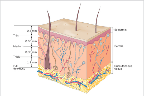
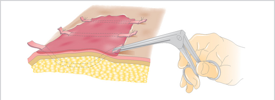
7 Grafting is an effective method for the management of granulation tissue but is not usually suitable for managing cases where there are identifiable reasons for the non-healing of the wound14. If the wound is affected by chronic and deep-seated infection or has foreign bodies, sarcoid cells, excessive movement, poor blood supply, an inappropriate pH for healing, or necrotic tissue or impaired blood supply it is unlikely to heal with grafts15. Free skin grafts should be considered in situations when there is a full thickness skin deficits, epithelialization is not active or is retarded, and when wound contraction is not occurring. Grafting should also be considered when conventional suturing techniques and sliding flaps are not possible; large defects below the carpus and hock frequently fall into this category. Spontaneous healing in these cases will be protracted and often results finally in dense (cheloid or hypertrophic) scar (see p. 89). 1. Autograft: tissue is taken from the animal itself. 2. Allograft (homograft): tissue is taken from the same species but a different animal. 3. Xenograft (heterograft): tissue is taken from a different species. At least one attachment to the donor site is maintained during healing. Flaps of skin with a broad attachment can sometimes be used to cover difficult wound sites (e.g. eyelid injuries). In some locations it may be possible to use skin stretching (balloon) systems before attempting to perform a pedicle graft. The commonest form of pedicle graft in horses is conjunctival grafting for corneal injuries and ulcerations (Figure 62). There are various forms of flap graft that can be used, including Y- and Z-plasty and tube grafts. These are described in surgical texts. The donor skin is dependent from the outset on the recipient site for its nutrition. There are two main forms that are simply classified in terms of the thickness of the skin graft, and therefore on the extent of adnexal structures. The thinner grafts (split thickness grafts) have no hair follicles, while the thicker ones (full thickness grafts) have intact hair follicles (Figure 63). Figure 63 Drawing of skin showing the position of the sectioning of skin for the various skin grafting techniques. (Modified from JA Auer and JA Stick, Equine Surgery, 2nd edn, 1999, WB Saunders.) ‘Postage stamp’ grafts (modified Meek method) uses small squares of skin (usually around 3–5 mm square) attached to an adhesive dressing. A special machine is used for preparation of the squares but simply cutting the skin into small squares could in theory produce suitable donor skin. The method allows the further expansion of the donor area to 1.5–2 times the original. The grafts are not dependent on the survival of all the squares: if a few do not survive they do not affect the others. Cosmetically the results are excellent, but the major disadvantage is the need to ensure the grafts are immobilized. To this end a rigid limb cast is usually applied16. Tunnel (strip) grafts can be used when the graft bed is less than ideal. The cosmetic effects are inferior to mesh grafting but the technique is more practical17. It requires less time, effort and expertise, and can be performed with minimal equipment in the standing animal. Success is not usually the all or nothing phenomenon associated with mesh grafts. Narrow strips of donor skin are obtained by parallel incisions 2 mm apart (Figure 64). All subcutaneous tissue is removed with a scalpel. About four or five strips can be obtained from a single site, which is then closed with sutures. The grafts are placed using 8 cm-long alligator forceps with a 2 mm diameter. Starting at the periphery of the wound, the forceps are inserted 5–10 mm deep into the granulation tissue and then passed horizontally through it to emerge on the opposite side. The grafts are drawn through the newly created tunnel. Care is taken not to twist them. The exposed ends are sutured or glued to the skin at the wound margin. Figure 64 Drawing of the technique for tunnel grafting. In most cases there is no need to bring the grafts to the surface in the middle of the grafted field, but this can help if the granulation tissue is on a curvature.
Skin Grafting
Classification of Grafts
Pedicle Graft
Free Grafts
Full Thickness Grafts
Tunnel (Strip) Grafts
![]()
Stay updated, free articles. Join our Telegram channel

Full access? Get Clinical Tree


Skin Grafting
 . Classification of Grafts
 . Pedicle Graft
 . Free Grafts
 . Clinico-pathological Consequences of Grafting
 . Graft Take and Causes of Failure
 . Summary
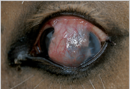
Only gold members can continue reading. Log In or Register to continue
