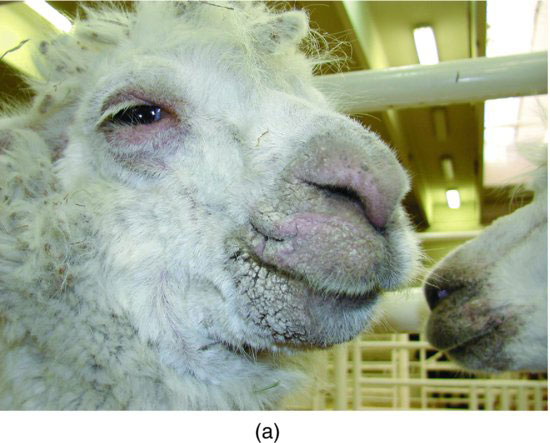
Figure 30.2 Alpaca affected by Sarcoptes. Sarcoptes can be difficult to confirm on skin scraping, but mites may be demonstrated on histopathology.
EQUIPMENT NEEDED
Clippers, exam gloves, biopsy punch, 20-gauge needle, syringe, 1 mL 2% lidocaine, 6- to 8-mm-diameter punch biopsy instrument, scissor or scalpel blade, specimen container with and without formalin, thumb forceps, and 2–0 skin suture and needle drivers or skin stapler will be needed.
RESTRAINT/POSITION
Standing, haltered, and chute restraint positions may be used.
TECHNICAL DESCRIPTION OF PROCEDURE/METHOD
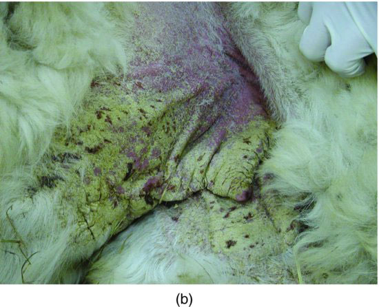
If the area to be sampled has fiber, clip the region without scraping skin, in order to maintain the integrity of the skin surface for histopathology. The skin surface should not be scrubbed or prepared with antiseptic. (If a sample is to be taken aseptically for bacterial culture, a second region should be aseptically prepared and the biopsy performed.) One mL of 2% lidocaine is injected subcutaneously by introducing the needle 8–10 mm away from the proposed biopsy site and injecting the lidocaine beneath the proposed site. If there is a discrete skin lesion, the biopsy site should be at the junction of normal/abnormal skin. Some histopathologists advocate placing the lidocaine remote to the skin being biopsied because lidocaine can cause vasodilation and edema in the sample. Alternatives to injection of lidocaine include topical lidocaine gel or hypothermia (ice packing) to numb skin prior to biopsy. These are somewhat less effective at achieving analgesia.
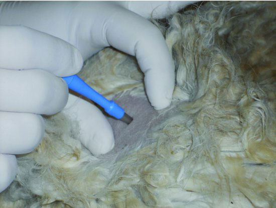
After approximately 5 minutes for lidocaine onset of action, the skin is stretched using the thumb and forefinger, and the biopsy punch is centered perpendicularly over the desired site (Figure 30.3). The punch is then rotated back and forth to “drill” into the skin (Figure 30.4). A release will be felt when the punch has entered the subcutaneous space and incised fully through the skin (Figure 30.5). The punch is removed and the circular sample will remain. The edge of the sample is gently grasped with thumb forceps (Figure 30.6) so the cellular structure of the sample is not crushed or altered. The sample is then lifted, and the subcutaneous connection is cut with Metzenbaum scissors or a scalpel blade (Figure 30.7).
Figure 30.3 Initial insertion of skin biopsy punch with slight skin stretching to facilitate the procedure.
Stay updated, free articles. Join our Telegram channel

Full access? Get Clinical Tree


