Chapter 103 Scapulohumeral Luxation
Scapulohumeral luxation is an uncommon clinical condition. Both congenital and traumatic luxations occur; the latter is more common.
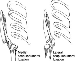
Medial luxation of the humeral head is most common, especially in smaller-breed dogs. Lateral luxation of the humeral head, while less common, usually occurs in larger dogs (Fig. 103-1). Cranial and caudal luxations are rare.
ANATOMY
• The shoulder joint is a ball-and-socket joint. The articular surface of the scapula forms the concave glenoid, and the convex humeral head articulates within it. Although the joint is capable of movement in any direction, its major actions are flexion and extension.
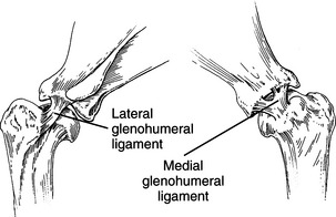
• The joint capsule is continuous and blends with the medial and lateral glenohumeral ligaments (Fig. 103-2). These ligaments help to support the joint and may be 2.0 mm or greater in thickness.
• The subscapularis, supraspinatus, infraspinatus, and teres minor muscles provide periarticular support.
• The tendon of origin of the biceps brachii and its sheath join with the joint capsule on its cranial medial aspect and provide additional stability. The bicipital tendon is held in the bicipital groove by the small but sturdy transverse humeral ligament.
DIAGNOSIS
• Careful palpation of the area between the acromion of the scapula and greater tubercle can be helpful.
PROCEDURES FOR CLOSED REDUCTION
Procedure for Medial Luxation
1. Flex the elbow and pull the limb laterally while exerting digital pressure on the scapular spine.
Stay updated, free articles. Join our Telegram channel

Full access? Get Clinical Tree


