40 SAMPLE COLLECTION AND PREPARATION
1 Why is cytology performed?
Cytology allows for the rapid determination of a diagnosis or identification of a process so that appropriate therapy can be provided quickly and cost effectively. Cytology is safe, with only minimal risks in most cases. Cytology is not a replacement for histopathology, but rather a complementary procedure.
2 Should the smears for cytology be stained or fixed before submitting them to the laboratory?
Generally, smears need not be stained and require no other special preparation before submitting them to the laboratory. Because most cytopathologists use a Romanowsky-type stain, the smears are simply allowed to air-dry and then packaged well to prevent breakage. There is no need to “fix” the smears in alcohol or other preservatives before mailing. Most laboratories want some if not all smears unstained in case special stains are needed, and many cytopathologists prefer using the stain they are most familiar with and staining the smears at the laboratory. If specific instructions are needed, however, the laboratory or the cytopathologist who will analyze the sample should be contacted with any questions.
3 What important considerations should one remember when mailing cytology smears?
A major problem is breakage in transit, so one should be sure to package the smears well. When submitting fluid samples, one should be sure to send the fluid in an EDTA tube and to send some premade smears from the fluid. Using premade air-dried smears of the fluid preserves the cells so that they can be evaluated better. Smears made from the fluid after arrival at the laboratory can have many artifacts in the cells that occurred in transit. The names of the owner and patient and the location where the sample was collected must be labeled in pencil on the frosted end of the slide to help prevent samples from being mixed up. Also, unstained cytology smears should never be mailed with histopathology samples. Formalin fumes fix the cells on cytology smears and inhibit their staining with Romanowsky stains and may make them uninterpretable.
4 Is special preparation of the collection site needed?
For cutaneous masses the site is prepared as is done for an injection. If a microbiologic culture is to be performed on the sample or if a body cavity is to be entered (e.g., abdominal, thoracic, or pericardial cavities; joints; transtracheal washes through cricothyroid ligament), the area of needle insertion should have a surgical scrub preformed.
6 Which size of needle and syringe should be used with the fine-needle technique (aspiration and nonaspiration)?
A small-gauge needle, between 25 and 22 gauge, should be used for fine-needle technique. Using a larger needle often results in more blood contamination and occasionally the collection of a core of tissue rather than individual cells.
Size of syringe is not a factor with the nonaspiration technique but varies with the aspiration technique. If one is aspirating a tissue that exfoliates cells easily, such as a lymph node, a smaller syringe (5 ml) can be used. For most tissues, however, a 12- or 20-ml syringe is needed to have sufficient negative pressure to collect tissue cells.
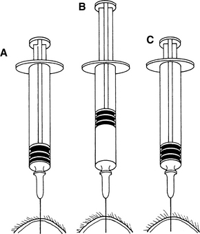
7 How is fine-needle aspiration performed?
For fine-needle aspiration of cutaneous masses, the mass is stabilized between the thumb and index finger of one hand while the needle (with syringe attached) is introduced into the mass. Negative pressure is applied by rapidly withdrawing the syringe plunger two-thirds to three-fourths the syringe volume. Multiple areas of the mass should be aspirated. If the mass is large enough and the animal calm enough, the needle may be redirected in the mass while negative pressure is maintained. If the mass is small or the animal difficult to restrain, the negative pressure should be released, the needle redirected, and negative pressure reapplied (Figure 40-1).

Figure 40-1 Fine-needle aspiration from a solid mass. After the needle is within the mass (A), negative pressure is placed on the syringe by rapidly withdrawing the plunger (B), usually half to three-fourths the volume of the syringe barrel. The needle is redirected several times while negative pressure is maintained, if done without the point leaving the mass. Before removing the needle from the mass, the plunger is released, relieving negative pressure on the syringe (C).
(From Cowell RL, Tyler RD, Meinkoth JH: Diagnostic cytology and hematology of the dog and cat, ed 2, St Louis, 1999, Mosby.)

Stay updated, free articles. Join our Telegram channel

Full access? Get Clinical Tree

