51 REPTILIAN HEMATOLOGY
2 What are some important principles to understand about the identification of reptilian leukocytes by blood smear evaluation?
3 What are some important considerations in evaluating leukocyte responses in reptiles?
4 Describe the various reptilian leukocytes.
The heterophil is the predominant leukocyte in health in some reptiles and predominates in many disease conditions, as discussed in the following questions. In most species of reptiles (chelonians, crocodilians, snakes, some lizards) the nucleus is round to oval, although the nucleus is lobulated in a few lizards (e.g., Green iguana). The heterophil of most reptile species has a cytoplasm that is densely packed with elongate, rod- to spindle-shaped, orange to brick-red granules that may partially obscure the nucleus (Figure 51-1). In other species the granules may not be as numerous.
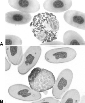
The eosinophil has prominent round, small cytoplasmic granules that vary in color from bright red to pink depending on the species (Figure 51-2). In a few species (e.g., Green iguana) the granules may appear lavender blue in color using Romanowsky-type stains. One must keep in mind that the granules of the eosinophil will stain differently than those of the heterophil within the same blood smear.
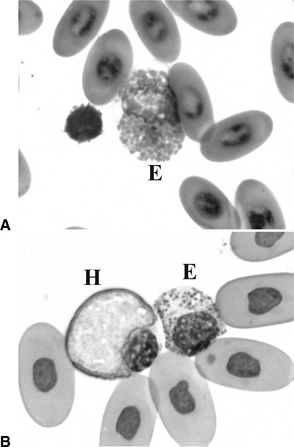
The basophil has prominent, small, deep-magenta to purple, round cytoplasmic granules and, unlike the mammalian basophil, has a round to oval, nonlobed nucleus (Figure 51-3). The contents of basophil granules can be dissolved by Romanowsky-type stains, giving the basophil a foamy, vacuolated appearance with few granules, if any. Careful examination of these cells in a blood smear may reveal a few characteristic granules, particularly over the nucleus, helping to identify them as basophils. Some species of reptiles (e.g., turtles) may have naturally high numbers of circulating basophils.
The monocyte is similar to its mammalian counterpart. It is a large mononuclear cell with abundant cytoplasm and a round to oval to indented nucleus (Figure 51-4). Cytoplasmic vacuolation may be seen. The azurophil is morphologically similar to the monocyte and is unique to some species of reptiles (snakes, iguanas). Some species of snakes in particular have greater numbers of azurophils than other reptiles. The azurophil is generally regarded as a cell of monocytic origin that contains numerous fine, red intracytoplasmic granules. The granules may be numerous enough to impart a red color to the cytoplasm.
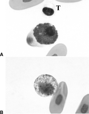
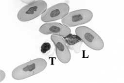
The lymphocyte is also morphologically similar to that of mammals. It is the predominant leukocyte in many species of reptiles (up to 80% of circulating lymphocytes). The lymphocyte is a mononuclear cell, with low to scant amounts of blue cytoplasm, high nuclear/cytoplasmic ratio, and a round nucleus (Figure 51-5). Reptiles normally have small and larger lymphocytes in circulation. Care must be taken not to confuse the small lymphocytes with thrombocytes. Thrombocytes may be smaller, more oval, with a darker, more condensed nucleus and indistinct, pale to nonstaining cytoplasm, and in contrast to lymphocytes, may readily clump in blood smears.
5 What are common methods of enumerating total numbers of leukocytes?
7 What causes heterophilia in reptiles?
Heterophilia in reptiles may be caused by the following two conditions, acting singly or in combination (Box 51-1):
Box 51-1 Causes of Heterophilia in Reptiles
11 When does hematopoietic tissue increase production of heterophil precursors in reptiles?
Increased hematopoietic production of heterophil precursors occurs when there is increased demand for heterophils (e.g., inflammation) in the peripheral tissues. Infection (pneumonia, enteritis, dermatitis), trauma, infarction, and neoplasms may result in increased demand for heterophils. Chemical mediators released from inflammatory cells, infectious agents, and damaged tissue stimulate the bone marrow to increase production. In healthy reptiles, hematopoietic production occurs primarily in the bone marrow. During conditions of increased demand, however, granulopoiesis may occur in extramedullary sites, such as the spleen, liver, and kidney.
12 What are the expected complete blood count findings in reptiles with heterophilia caused by corticosteroid effects?
13 What are the expected CBC findings in reptiles with heterophilia of inflammatory disease?
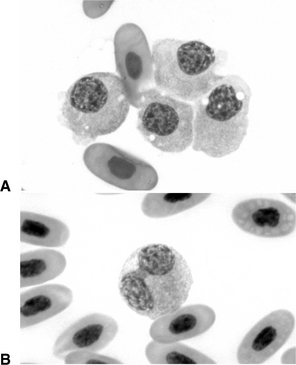
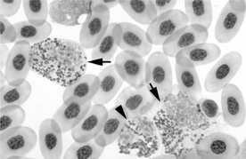
Mild heterophilia may be the only CBC abnormality in mild inflammatory disease. With increasing severity, left shift (increased presence of immature heterophils, e.g., band forms, metamyelocytes, myelocytes, promyelocytes; Figure 51-6), heterophil toxicity (cytoplasmic basophilia, abnormal vacuolation, abnormally shaped cytoplasmic granules, degranulation, large heterophils), and greater numbers of heterophils may be present and reflect a significant demand for heterophils in tissues. Monocytosis or azurophilia may also be observed. Overwhelming inflammation may result in heteropenia with left shift and heterophil toxicity (see Question 15). The number of lymphocytes may be decreased, normal, or increased with inflammatory disease. Environmental factors (e.g., temperature) may also influence the magnitude of the changes.
14 What are causes of heteropenia in reptiles?
Heteropenia may be caused primarily by overwhelming inflammation resulting in excessive tissue demand for heterophils (Box 51-2).
Box 51-2 Causes of Heteropenia in Reptiles
Stay updated, free articles. Join our Telegram channel

Full access? Get Clinical Tree


