CHAPTER 137 Reproductive Patterns in Female Bison (Bison bison sp.)
REPRODUCTIVE CHARACTERISTICS
The female bison is seasonally polyestrous, with estrous cyclicity usually beginning in late July or August and conception generally occurring at the first or second estrus and a reported 95% of cows mating only once.1–4 The length of the bison estrous cycle has been reported to range from 16 to 31 days, with an average length of 20.8, 21.5, and 20.8 days based on behavioral observations, and urine and fecal progestin profiles, respectively.2,3 Although estrogens can be measured in cycling female bison, the profiles do not provide physiologic indicators of behavioral estrous or ovarian activity.5 Based on endocrine evaluations of luteal activity, behavioral signs of estrus, and recorded births, females can continue to cycle through December or January.3,6 Demonstrations of behavioral estrus include homosexual mounting, standing for mounting, flehmen, and tail flagging, and these behaviors coincide with nadir progestin values in cycling animals.3,5,7 The behavioral ecology of male and female bison, with particular emphasis on reproductive behaviors, has been well described by Berger and Cunningham.4
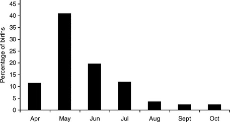
To establish population demographics and reproductive characteristics in bison, historical records from the Toronto Zoo wood bison herd were analyzed from 1978 to 2000.6 Of the 145 bison calves born during this time, all were singletons. A total of 14 calves (9.6%; 8 males, 4 females, and 2 unknown) either were stillborn or died on the day of birth. Eleven calves (7.6%; 3 males, 8 females) died within 30 days post partum. Parturition most frequently occurred in May (Fig. 137-1, 55.4%). This is consistent with data from free-ranging plains bison; however, animals living at higher latitudes or in areas with more harsh winters have demonstrated a trend to produce calves later in the spring.4 From the Toronto Zoo records, calves born from August to October had a greater mortality rate (37.5%), compared to those born between April and July (15.5%).6 The sex ratio of offspring was approximately equal: 51.0% females and 47.6% males. The youngest and oldest age at which a female produced offspring was 2 and 17 years, respectively. The mean age of females producing offspring (from females of known age, n = 102) was 6.01 years. On average, both bulls and cows lived between 14 and 17 years.
Calf production in free-ranging plains bison has been reported to be greatest in females aged 5 to 13, with those aged 10 to 13 years being the most fecund, and more than 80% of pregnancies occurring in animals 4 years of age or older.4,8 Pregnancy rates have been reported in lactating cows and are attributed to ovulation failure.8 Although rare, yearlings have been reported to conceive.1,4 Pregnancy rates of 70% have been reported for free-ranging bison.1 Observations of 16 plains bison births have shown that parturition is rapid, with birth occurring an average of 68 minutes after the amnion was first visible and 13 minutes after the forehooves were first observed.9 Calves were precocial and stood and nursed 10.8 and 32.2 minutes, respectively, after birth. In open habitat, cows tended to remain in groups, whereas in wooded areas, cows tended to give birth in isolation.9
PREGNANCY
There are no previous reports documenting hormonal patterns during the entire period of gestation in bison. To establish longitudinal steroid profiles for the wood bison during pregnancy, fecal and urine sample collection was performed at least five times weekly over three reproductive seasons (August until birth) from a captive herd of wood bison at the Toronto Zoo. The actual number of samples for each individual varied, but samples were taken at least once each week. Samples were analyzed for steroid hormone concentrations by enzyme immunoassay (EIA) using previously described methods.3,5,7 Urine was analyzed for pregnanediol-glucuronide (PdG), estrone conjugates (EC), and cortisol, and feces were analyzed for progestins (P) and EC. Steroid hormones were extracted from feces according to previously described methods and stored frozen until analysis.5,7 Gestation length was determined from observed mating or last observed estrus until the birth of a calf.
Hormonal patterns were monitored in 11 pregnancies over the 3-year period. One calf from year 1, one calf from year 2, and two calves from year 3 were stillborn, and their respective dams died due to complications during or after calving. Two of the stillbirths were female and two were male. The mean gestation length for the 11 pregnancies was 265.3 ± 2.5 days. This differs from reports of primiparous and multiparous free-ranging bison (287.9 versus 279.9, respectively). Berger and Cunningham have reported adjustment of gestation length in females, with those in good body condition experiencing an ∼6 day shortening of gestation.4 No difference was detected between pregnancies producing live offspring (267.9 ± 1.0 days) and stillbirths (263.2 ± 6.8 days) or between pregnancies carrying male and female calves in the Toronto Zoo wood bison herd. Physical attributes indicative of impending parturition included: vulval swelling 2 to 3 weeks prepartum, mucus secretion approximately 2 weeks prepartum, and udder development. On the day of or just preceding parturition, increased restlessness, separation from the herd, passing of the mucous plug, and contractions were observed.5
Live Births
Progestins
Composite weekly PdG and P profiles from the viable pregnancies are shown in Figure 137-2. Urinary PdG concentrations were unchanged from week −37 until week −14, then increased markedly from week −13 to week −7. After week −7, PdG levels declined gradually to 749.0 ± 148.1 ng/mg creatinine at week −5, followed by a more dramatic decrease at week −4, which continued through week 0. Fecal P levels demonstrated a pattern similar to the PdG profile (see Fig. 138-2). Mean P concentrations did not differ from luteal phase values between week −37 and week −12. A significant elevation in P was detected at week −10 (920.56 ± 124.6 ng/g feces). Fecal P values then continued to rise to a peak at week −4, after which time levels markedly declined through week −2 and continued to fall through parturition. Fecal P concentrations had not reached baseline by week 3 post partum.
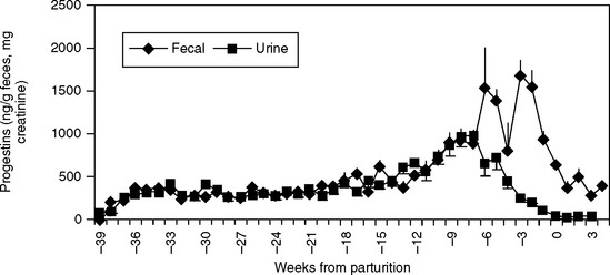
 ) concentrations during gestation in seven wood bison at the Toronto Zoo. Data are aligned to the week of parturition (week 0).
Estrone Conjugates
A doubling in mean urinary EC from mean nonpregnancy levels of 1.0 ± 0.1 ng/mg Cr to 2.2 ng/mg Cr was detected between weeks −35 to −33 (Fig. 137-3). Urinary EC then remained statistically unchanged, but increased gradually, from week −31 to week −5, then increased rapidly between week −4 to a peak at week −1 (range: 475.2–2272.7 ng/mg Cr). A precipitous decline in urinary EC was observed between week −1 to week 0 and decreased even further by week 3 post partum. Fecal EC concentrations increased markedly from mean nonpregnancy levels (25.4 ± 5.7 ng/g feces) between weeks −30 to −26 (see Fig. 137-3), similar to the pattern observed for urinary EC. Fecal EC concentrations remained statistically unchanged from week −30 to week −5, but also showed a gradual increase over time. A significant elevation in fecal EC concentrations was detected at week −4 (295.5 ± 29.9 ng/g feces) which increased in all females to a peak during week −1. Fecal EC concentrations declined in the week prior to parturition and continued to fall through week 3.
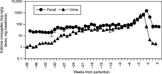
 ) estrone conjugate concentrations during gestation in seven wood bison at the Toronto Zoo. Data are aligned to the week of parturition (week 0).
Stay updated, free articles. Join our Telegram channel

Full access? Get Clinical Tree


