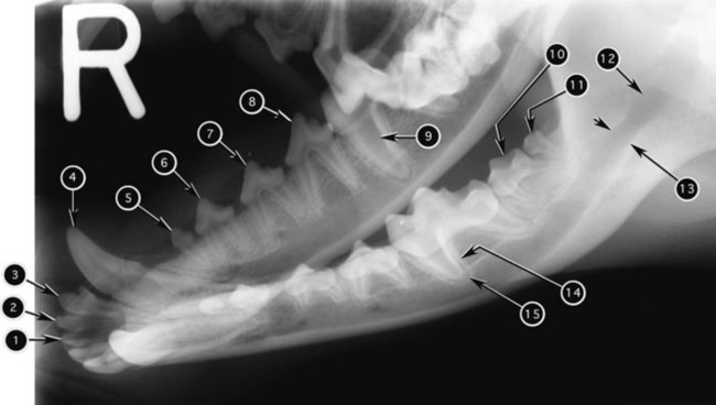
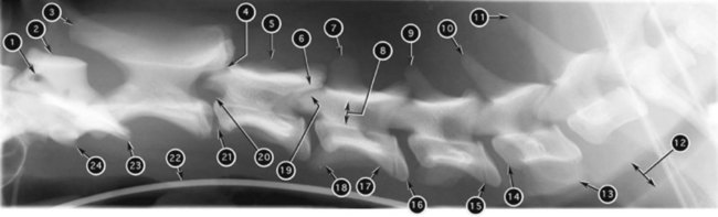
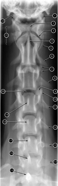
To use the roentgen sign method of recognizing abnormal radiographic findings effectively, an understanding of normal radiographic anatomy for the specific area of interest is necessary. Within the space constraints of a comprehensive veterinary radiology text, this chapter provides a limited reference for the radiographic anatomy of the axial skeleton. For more detailed information, readers are referred to comprehensive texts on radiographic anatomy.1,2,2 The radiographic nomenclature used in this chapter was approved by the American College of Veterinary Radiology in 1983.4 1. Lateral vertebral foramina (left and right) of atlas; emergence of cervical nerve 1 3. Spinous process of axis (C2) 4. Synovial joints between articular processes of C2 and C3 6. Caudal articular processes of C3 13. Expanded ventral laminae of transverse processes of C6 14. Cranial extremity (head) of C6 16. Caudal extremity (fossa) of C4 18. Transverse processes of C4 19. Cranial articular processes of C4 20. Intervertebral foramina between C2 and C3 21. Intervertebral space (disk) between C2 and C3 23. Wings (transverse processes) of atlas 6. Left transverse process of axis 8. Left transverse process of C3 9. Left caudal articular process of C3 10. Left cranial articular process of C4 11. Left transverse process of C4 12. Left transverse process of C5 13. Left transverse process of C6 14. Left transverse process of C7
Radiographic Anatomy of the Axial Skeleton
![]()
Stay updated, free articles. Join our Telegram channel

Full access? Get Clinical Tree


Radiographic Anatomy of the Axial Skeleton