Pythiosis
Basic Information 
Definition
Deep, invasive, and rapidly progressive subcutaneous oomycotic infection caused Pythium insidiosum
Clinical Presentation
Physical Exam Findings
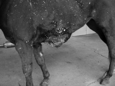
• P. insidiosum generally infects the wounds in the distal portions of the body, including the limbs below the carpus and hock, ventral abdomen (Figure 1), and ventral thorax (areas more likely to be in contact with stagnant water), as well as the lips, nostrils, face, external genitalia, neck, and trunk.
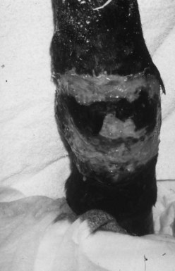
 Lesions are usually single and large nodular eroded to ulcerative granulomas associated with moderate to severe pruritus and oozing of serosanguineous discharge (Figure 2).
 If excised, tissue has a thickened fibrotic dermis (pink) and contains scattered areas of red surrounding a yellow-tan, gritty, central necrotic core (“leeches” or “kunkers”). Kunkers range from 2 to 10 mm, which is larger than those seen with zygomycosis.
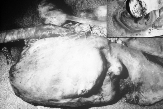
 Lesions involving the limbs are accompanied by lymphangitis with severe edema or swelling (Figure 3)
 The infection is locally invasive (spreading from the subcutaneous to lymphatics and deeper tissues (eg, bones, tendon sheaths, joints).
• Lesions are often heavily contaminated by bacteria (especially gram-negative organisms).
< div class='tao-gold-member'>
Only gold members can continue reading. Log In or Register to continue
Stay updated, free articles. Join our Telegram channel

Full access? Get Clinical Tree


