Figure 54.2 The rigid sigmoidoscope has an obturator that is blunt ended making easier entry through the vulva and into the vagina.
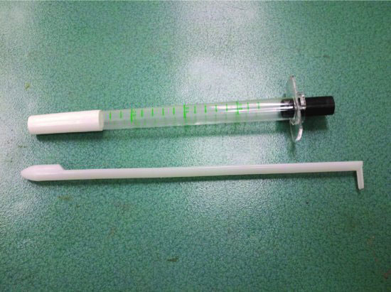
Figure 54.3 The sigmoidoscope light adaptor from Welch-Allyn can be attached to most standard hand-held power supplies used for otoscopic and ophthalmic examinations.
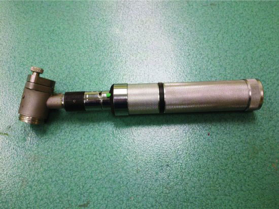
Figure 54.4 The sigmoidoscope light adaptor provides a magnification lens and circumferential light to facilitate examination without light source interference.
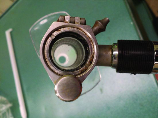
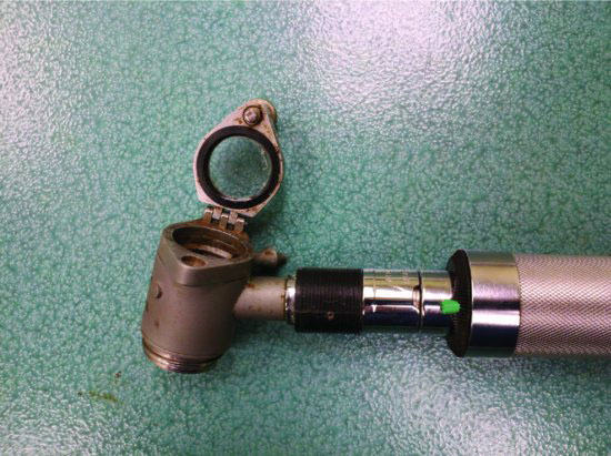
Figure 54.5 The magnifying lens may be opened to allow for sample collection when cytology or cultures are desired.
Insertion of the vaginoscope is done by applying sterile lubricant to the exterior of one end of the scope. Then, the scope is inserted through the vulva at a 60-degree ventral angle to the spine (the scope will be partially vertically oriented). At this angle, the scope is pushed dorsal and cranial for 5 to 6 cm until slight resistance is felt. When resistance is felt, the free end of the vaginoscope is elevated until advancement is possible. The final position of the vaginoscope is approximately horizontal. The light source is attached or inserted and the cervix identified and inspected (Figure 54.6). The cervix is recognized by the spiral form of the external os. The cranial vagina and cervix normally have a pink color and smooth surface with an absence of fluid. In the presence of a dominant follicle, the cervix is relaxed (“open”) and can be recognized by the presence of a noticeable lumen. In the presence of a CL, the cervix is “closed,” and the lumen is difficult to detect.
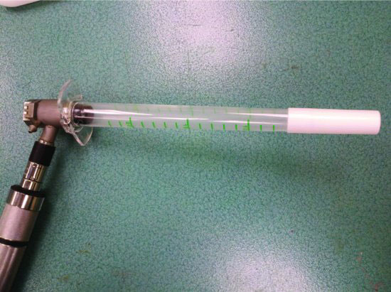
Figure 54.6 The light source adaptor is attached to the sigmoidoscope by threads. This allows the unit to be held and manipulated as one piece with one hand.
Stay updated, free articles. Join our Telegram channel

Full access? Get Clinical Tree


