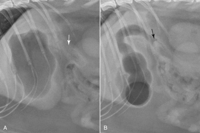
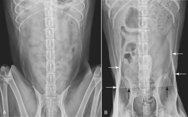
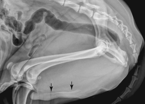
As with the thorax, the naming of lateral abdominal radiographs made with the patient recumbent, as commonly done in dogs and cats, violates the point-of-entrance to point-of-exit nomenclature system described in Chapter 5. If that system were followed, a lateral view made with the dog or cat in left recumbency would be termed a right-left view because the x-ray beam strikes the right aspect of the abdomen and exits the left aspect of the abdomen. But, for the abdomen, the terminology for lateral projections has been abbreviated to describe the side of the patient lying on the x-ray table. Thus, for example, an abdominal radiograph acquired with a dog or cat lying on the left side would be termed a left lateral. A lateral and a VD radiograph will be adequate for assessing the abdomen completely in many canine and feline patients. However, the abdomen contains a valuable inherent contrast medium—gas. Thus, by routinely acquiring left and right lateral radiographs in addition to the VD view, redistribution of bowel gas between the two lateral views can provide valuable additional information that can make the difference between obtaining the diagnosis or not (Fig. 35-1). Dorsoventral radiographs of the abdomen are rarely made and should not form the basis of the orthogonal view to complement the lateral projections. A DV view may have to suffice when a patient cannot be positioned in dorsal recumbency for a VD view, but the abdomen will be more crowded, organ conspicuity will be reduced, and portions of the pelvic limbs are often superimposed on the abdomen (see Fig. 35-9 for an example). It is important that the entire abdominal cavity be included in the image. The field of view should extend from just cranial to the diaphragm to a few centimeters caudal to the coxofemoral joints. For lateral views, the pelvic limbs should not be pulled caudally but should be kept perpendicular to the spine. This relaxes the caudoventral aspect of the abdominal wall and reduces crowding. Likewise, in the VD view, the pelvic limbs should be flexed into a so-called frog-leg position rather than being pulled caudally. If the pelvic limbs are pulled caudally for the VD view, crowding will be increased, and skin folds will be created in the lateral thigh and/or caudal abdomen region. These skin folds create noticeable superimposed opacities that will interfere with assessment of the caudal aspect of the abdomen (Fig. 35-2). Right and left lateral and VD views of the abdomen will suffice for almost all canine and feline patients. If the status of the urethra in male dogs is of concern, as for assessment of urethral calculi, the right and left lateral views should be supplemented with a third lateral view made with the pelvic limbs pulled cranially. This provides an unobstructed view of the ischial arch and os penis, allowing for assessment of urethral calculi without superimposition of the pelvic limbs (Fig. 35-3). A compression radiograph can be used to clarify a suspicious finding that cannot be confirmed because of superimposition.1 The theory is to apply mild compression to displace superimposed organs from the field of view, leaving an unobstructed view of the region of interest. This technique can be applied to assess structures such as the uterine body (lateral view) or the caudal aspect of the retroperitoneal space (VD view). Specialized inflatable compression paddles are available, but a wooden spatula or wooden spoon can also be used. If using a film-screen system, the kVp should be decreased by approximately 15% because the compressed part will be reduced in thickness. Of course, care should be taken not to place compression on organs that are obviously diseased, and the amount of pressure placed should always be gentle (Fig. 35-4). The increased use of ultrasound for abdominal assessment has reduced the need for compression radiographs, but they remain a convenient method to increase the accuracy of radiographic interpretation in some patients where ultrasonography is not possible. One major difference between left and right lateral abdominal radiographs in dogs and cats is the appearance of the stomach. This relates to the difference in the anatomic position of the gastric fundus versus the gastric pylorus.2,3 When looking at a cross-section of the abdomen from the caudal aspect of a dog or cat, the fundus is located dorsally and on the left versus the pylorus, which is located ventrally and on the right (Fig. 35-5
Principles of Radiographic Interpretation of the Abdomen
Nomenclature
Positioning—Dog and Cat
Lateral View
![]()
Stay updated, free articles. Join our Telegram channel

Full access? Get Clinical Tree


Principles of Radiographic Interpretation of the Abdomen