If the female is bred on day 0 and has a progesterone level >2.0 ng/mL on day 7, the interpretation is that she ovulated. If the progesterone remains increased at day 14, the interpretation is that she conceived a pregnancy. Alternatively, she may have not conceived but rather developed a retained CL. Increased progesterone on day 21 is interpreted as successful implantation of the pregnancy into the uterus. Alternatively, the female could have a retained CL, luteinized follicle or other progesterone-secreting tissue.
- Inappropriate increase in progesterone
- Persistent CL
- Cystic CL
- Ovarian tumors
- Persistent CL
- Granulosa cell tumor
- Granulosa theca cell tumor
Ultrasonography: Ultrasonography is the preferred method of diagnosis of pregnancy in llamas and alpacas.
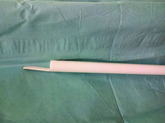
Transrectal ultrasonography is done most commonly via rectal examination. The small size of camelids limits the examiner’s ability to perform rectal palpation for control of the linear probe. A rigid extender can be manufactured from PVC pipe that is molded to the contour of a linear probe to facilitate control of the probe during the examination (Figure 53.1 and Figure 53.2). This must be done with extreme care so as not to injure or tear the rectum. Ideally, the camelid is restrained in a chute so that movements are restricted. Sedation is advisable to prevent or minimize unwanted activity during the examination. A lubricant (60 to 120 mL) should be instilled into the rectum prior to insertion of the probe. The nongravid uterus is recognized as a homogeneous gray to white tissue approximately 1 cm in diameter (Figure 53.3). The uterine horns are often curled in a “C” shape, and the ovaries can be found cranial and lateral to the tip of the nongravid uterus (Figure 53.4). The gravid horns straighten and extend cranially with advancement of the pregnancy. Pregnancies <45 days are most easily examined via transrectal ultrasonography (Figures 53.5 to 53.7). Pregnancies >60 days but <180 days are most easily found via transabdominal ultrasound on the left side of the abdomen approximately 10-cm cranial to the brim of the pelvis and medial to the stifle (Figures 53.7 and 53.8). Pregnancies >180 days are difficult to find by ultrasound examination. At this stage, the cria resides cranial, ventral, and to the right side of midline in the abdomen. The cria thorax is most consistently found 10- to 20-cm caudal to the xyphoid and 10- to 15-cm to the right of ventral midline (Figures 53.9 to 53.12).
- Transrectal
- <45 days
- Up to 90 days
- <45 days
- Transabdominal
- Left flank
- 60 to 180 days
- Right paramedian
- >180 days
- Left flank
Figure 53.1 A rigid extension can be made from rigid plastic or polymer tubing to facilitate transrectal ultrasonography when transrectal palpation is not possible. Extreme care must be exercised to prevent tearing or other damage to the rectum.

Stay updated, free articles. Join our Telegram channel

Full access? Get Clinical Tree


