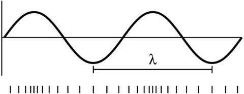
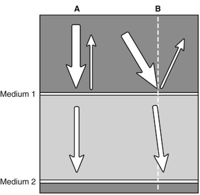
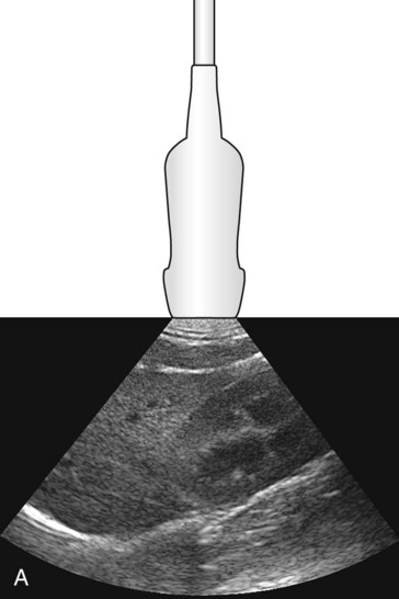
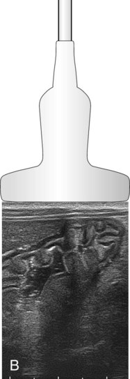
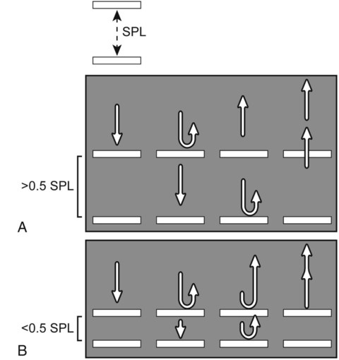
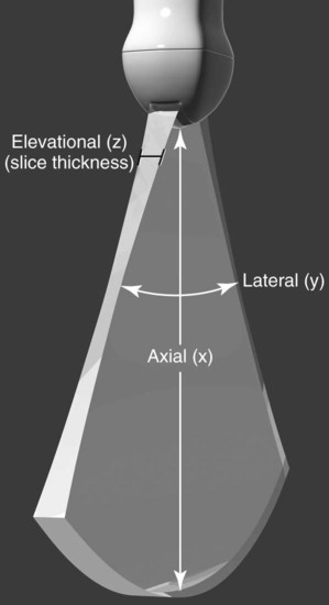
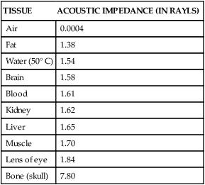
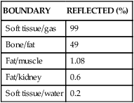
Sound travels in waves and carries information from one location to another. It transmits energy by alternating regions of low pressure (rarefaction) and high pressure (compression).1–4 Unlike light and radio waves, sound waves require a medium through which to travel.5 Frequency, wavelength, and velocity are parameters used to describe sound waves; these terms are also used in reference to electromagnetic radiation (see Chapter 1). Frequency is the number of times a wave is repeated per second. One wave or cycle occurs when pressure starts at a normal value, increases to a high value, decreases (passing the normal value) to a low value, and then returns to normal (Fig. 3-1). A cycle may also be defined as the combination of compression and successive rarefaction.1 Frequency is expressed in hertz (Hz), where 1 Hz equals one cycle per second. In diagnostic ultrasound, the frequencies are typically between 2 megahertz (MHz) and 15 MHz (1 MHz = 1,000,000 Hz). The audible range of sound frequencies for human beings is 20 Hz to 20,000 Hz; sound less than 20 Hz is infrasound, and sound greater than 20,000 Hz (0.02 MHz) is ultrasound.1,4,4 Velocity is the rate at which sound travels through an acoustic medium; it is determined by the physical density (mass per unit volume) and stiffness (hardness) of the transmitting medium.1,2 The velocity of sound in some commonly encountered tissues is listed in Table 3-1. If physical density remains constant, velocity increases as stiffness increases. If the stiffness remains constant, velocity decreases as physical density increases. As a general rule, velocity is highest in solids, lower in liquids, and the lowest in gases. In solids, molecules are closer together, so sound waves are transmitted faster; in gases, the molecules are far apart, and sound waves travel more slowly. Medically, sound waves travel the fastest in bone and the slowest in gas-filled lungs. Velocity is related to frequency and wavelength of a sound wave in the following equation: Table • 3-1 Velocity of Sound in Commonly Encountered Tissues2 For a constant velocity, frequency and wavelength have an inverse relationship so that as frequency increases, wavelength decreases, and vice versa. Within soft tissue, the average velocity of sound is 1.54 mm/µs (1540 m/s).1–5 The velocity of sound in soft tissue is important because ultrasound machines use this constant velocity for all calculations. Acoustic impedance of a tissue is the product of the tissue’s physical density and sound velocity within the tissue.1,4 Changes in acoustic impedance from one tissue to another determine how much of the sound wave is reflected and how much is transmitted into the second tissue. The amplitude of the returning echo is proportional to the difference in acoustic impedance. If two tissues have no difference in acoustic impedance, then no echo is created. If a large difference in acoustic impedance exists between two tissues, then almost all the sound is reflected in an echo.1,2 The following formulas are used to calculate the percentage of the sound wave that is reflected and transmitted:1,3,3 In Equation 3, Z2 is the acoustic impedance of the second tissue, and Z1 is the acoustic impedance of the first tissue. The approximate acoustic impedance of tissues encountered commonly is listed in Table 3-2 and the percentage of sound reflected at various tissue interfaces is listed in Table 3-3. By the values in Table 3-2, the largest difference in acoustic impedance occurs at interfaces with bone and gas. Almost all sound is reflected at soft tissue/gas and soft tissue/bone interfaces. Thus little or no sound passes these boundaries. Hence the speed of sound in bone and gas-filled lung is not important clinically. This nearly total sound reflection creates an acoustic void, or shadow, deep to the tissue interface. Without placement of acoustic coupling gel between the patient and transducer, the soft tissue/gas interface created by gas trapped between the skin and transducer would prevent imaging because of reflection of all oncoming sound. Table • 3-2 Approximate Acoustic Impedance in Commonly Encountered Tissues2 Table • 3-3 Percentage of Sound Reflected at Interfaces5 Angle of incidence is the angle at which a sound wave encounters a medium. If the angle of incidence is perpendicular (90 degrees) to the reflector, the reflected portion of the sound wave travels 180 degrees opposite to the direction of the initial sound wave, and the transmitted portion of the sound wave continues in the same direction as the initial sound wave (Fig. 3-2, A). If the angle of incidence is not perpendicular, then the angle of reflectance equals the angle of incidence (Fig. 3-2, B). If a sound wave strikes a reflector more than 3 degrees from perpendicular, then the reflected sound will likely not reach the transducer.2 The angle of transmission of a nonperpendicular sound wave depends on the relative acoustic impedance of the two media. Any sound wave that is not reflected toward the transducer will not be recorded. A key fact is that the amount of reflected and transmitted sound depends on the difference in acoustic impedance of the two media. An ultrasound beam is attenuated, or loses strength, as it travels through a medium. The amount of attenuation is determined by the distance traveled and the frequency of the sound wave. The amount of attenuation approximates 0.5 decibels (dB)/cm per MHz over a round-trip distance.4 If a reflector is 3 cm from the transducer, the round trip distance is 6 cm, and the attenuation of the sound beam is 3 dB/MHz. High-frequency sound waves are attenuated more than are lower-frequency sound waves. Attenuation of the sound wave involves three components: absorption, reflection, and scattering.1–3 Absorption is the conversion of the mechanical energy of a sound waves to heat.1,3 Absorption is the dominant component of attenuation of sound in soft tissue.1,3 The conversion of sound waves to heat occurs primarily by frictional forces.3,4 With diagnostic ultrasound machines, the relative amount of sound absorbed is quite low, and the temperature change is insignificant and imperceptible. Scattering occurs when sound waves encounter small, irregular surfaces. This occurs mainly within the parenchyma of organs and is responsible for the echotexture of internal organs.1,3 As the frequency of a sound beam increases, the incidence of scattering increases. A transducer is a device that converts one form of energy to another.3,6 In ultrasound imaging, transducers convert electric current into sound waves, and vice versa.7 This conversion is accomplished in a piezoelectric crystal. As an electric charge is applied to a piezoelectric crystal, the material deforms and creates a sound wave.6 Conversely, when sound waves are applied to piezoelectric crystals, they produce an electric signal. Therefore the same crystal is used to send and receive sound waves, but these two processes cannot occur at the same time.1 For typical ultrasound imaging, the transducer emits sound waves less than 1% of the time and receives sound waves more than 99% of the time.4 Electronic transducers, also called array transducers, are composed of several small elements in various arrangements. The elements may be arranged in a line or rectangle (linear array), in a curved line (convex array), or in concentric ring fashion (annular array).8 The elements that compose these transducers are electronically fired in various sequences to create different-shaped images or to focus the sound beam at specific depths.7 Two basic shapes of ultrasound images are commonly encountered. Images made in sector fashion are pie shaped (Fig. 3-3, A), and images made in linear fashion are rectangular (Fig. 3-3, B). The footprint of a transducer is the amount of the transducer that makes contact with the patient. Sector images are often produced from transducers with a small footprint whereas linear transducers usually have larger footprints. For thoracic imaging, sector-shaped transducers are preferable because images must be acquired intercostally. For abdominal imaging, the use of sector or linear transducers is often dictated by the personal preference of the sonographer. In most instances, more than one transducer is used during a single examination. Diagnostic ultrasound machines operate in pulsed mode for imaging. This means that the ultrasound machine sends only a few cycles of sound into tissue and then spends the rest of the time listening for returning echoes. The pulse repetition frequency (PRF) is the number of times this pattern of sending and listening is repeated within 1 second.1 The length of space in one pulse of ultrasound is called the spatial pulse length (SPL). If a sound wave has a wavelength of 0.5 mm and three pulses are sent each time, the SPL is 1.5 mm. Spatial pulse length is important for axial resolution. Resolution is the ability of an ultrasound machine to distinguish echoes on the basis of space, time, and strength.6 The better the resolution, the more likely an abnormality can be identified. As the frequency of an ultrasound wave increases, the resolution increases. A transducer of the highest possible frequency should be used to get the best possible resolution. Axial resolution is the resolution of two separate reflectors along the direction in which the sound wave is traveling.6 It is equal to half the SPL. As previously mentioned, SPL is the length of space over which the pulse of a sound wave travels. When two reflectors are separated by a distance that is greater than half the SPL, the echoes from these two reflectors do not overlap as they return to the transducer and are interpreted as separate echoes (Fig. 3-4). If the distance between the reflectors is less than half the SPL, the returning echoes overlap and are interpreted as a single echo. Because transducers with higher frequency have a shorter SPL, axial resolution is improved. Lateral resolution is the resolution of two separate reflectors perpendicular to the direction in which the sound wave is traveling (Fig. 3-5).6 This is determined by the width of the ultrasound beam.6 To recognize the objects discretely, the ultrasound beam must be narrower than the distance between the objects. The width of an ultrasound beam decreases with increasing frequency. In a focused ultrasound beam, where the width of the beam is restricted, the lateral resolution is best at the focal point of the ultrasound beam because this is the narrowest part of the beam.6 Broad bandwidth technology allows for transducers to emit a wide range of frequencies. This allows the user to select various frequencies from the same transducer.6,7 With older narrow bandwidth transducers, a 4 MHz transducer might emit frequencies between 3.8 and 4.2 MHz, whereas a modern broadband 4MHz transducer can emit frequencies between 1 and 7 MHz.9 The broadband frequency transducers improve near-field resolution by using the higher frequency of the spectrum and far-field resolution by using the lower frequency of the spectrum. Additionally, axial resolution is improved because these transducers use shorter SPLs.9
Physics of Ultrasound Imaging
Physical Principles of Ultrasound Waves
TISSUE
VELOCITY (MM/µS)
Air
0.331
Fat
1.450
Water (50° C)
1.540
Soft tissue
1.540
Brain
1.541
Liver
1.549
Kidney
1.561
Blood
1.570
Muscle
1.585
Lens of eye
1.620
Bone (skull)
4.080
 (Eq 1)
Ultrasound Wave Interaction with Matter
 (Eq 2)
 (Eq 3)
 (Eq 4)
TISSUE
ACOUSTIC IMPEDANCE (IN RAYLS)
Air
0.0004
Fat
1.38
Water (50° C)
1.54
Brain
1.58
Blood
1.61
Kidney
1.62
Liver
1.65
Muscle
1.70
Lens of eye
1.84
Bone (skull)
7.80
BOUNDARY
REFLECTED (%)
Soft tissue/gas
99
Bone/fat
49
Fat/muscle
1.08
Fat/kidney
0.6
Soft tissue/water
0.2
Transducers
![]()
Stay updated, free articles. Join our Telegram channel

Full access? Get Clinical Tree


Veterian Key
Fastest Veterinary Medicine Insight Engine

