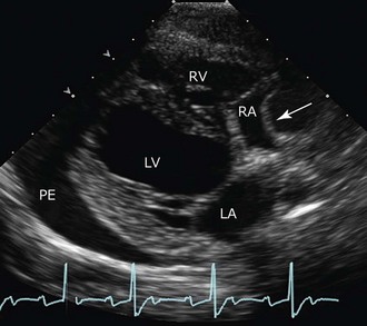
Pericardial Effusion

Chapter 182


Pericardial Effusion


The double-layered pericardial sac surrounding the heart normally contains a small volume (≈0.25 ml/kg) of serous fluid between its outer fibrous layer (parietal pericardium) and inner serous membrane (visceral pericardium or epicardium). Excessive fluid accumulation (pericardial effusion) is the most common pericardial disorder in small animals. It occurs much more frequently in dogs than in cats.


Pathophysiology


Pericardial effusion disturbs cardiac function by impeding filling. Because the pericardium is relatively noncompliant, increases in pericardial fluid volume can increase intrapericardial pressure sharply. When intrapericardial pressure equals or exceeds normal cardiac filling pressure, cardiac filling is impaired. This condition is known as cardiac tamponade. The rate of pericardial fluid accumulation and the distensibility of the pericardial sac determine whether and how quickly cardiac tamponade develops. Rapid accumulation of a small volume (e.g., 50 to 100 ml) can raise intrapericardial pressure markedly because the pericardium can stretch only slowly. Fibrosis and pericardial thickening may further limit the compliance of this tissue. Pericardial fibrosis and inflammatory cell infiltrates are seen with idiopathic and neoplastic causes of effusion. Conversely, if effusion accumulates slowly, the pericardium may enlarge sufficiently to accommodate the increased volume at low pressure. As long as intrapericardial pressure is lower than normal venous pressures, cardiac filling and output are maintained and signs of tamponade are absent. Therefore large-volume pericardial effusion implies a gradual process.


The external cardiac compression that occurs with tamponade progressively limits filling, initially of the right side of the heart but subsequently of the left side as well. Systemic venous pressure increases while cardiac output falls. Diastolic pressures in all cardiac chambers and great veins eventually equilibrate. Neurohumoral compensatory mechanisms of heart failure become activated as cardiac output falls. External signs of systemic venous congestion and right-sided congestive heart failure (CHF) become especially prominent with time.


Cardiac tamponade exaggerates the variation in arterial blood pressure that occurs normally during the respiratory cycle. If the inspiratory fall in left heart output causes a 10 mm Hg or greater fluctuation in systolic arterial pressure, this is known as pulsus paradoxus. Although myocardial contractility is not affected directly by pericardial effusion, inadequate ventricular filling combined with reduced coronary perfusion during tamponade can lead to systolic and diastolic ventricular impairment. Low cardiac output, arterial hypotension, systemic venous congestion, and poor perfusion of the heart, as well as other organs, can lead ultimately to cardiogenic shock and death.


Presentation


Cardiac tamponade is relatively common in dogs but rare in cats. Clinical signs reflect the consequences of systemic venous congestion and poor cardiac output. Right-sided congestive signs usually predominate, especially abdominal enlargement (ascites). Tachypnea and weakness or syncope with exertion are common in the history. Lethargy, poor exercise tolerance, inappetence, cough, and other nonspecific signs can occur before obvious ascites develops. Loss of lean body mass (cachexia) is apparent in some chronic cases. Rapid pericardial fluid accumulation can cause acute tamponade, shock, and death. In such cases clinical signs of jugular venous distention, hypotension, and possibly pulmonary edema may be evident without pleural effusion, ascites, or radiographic cardiomegaly.


Physical examination findings typically include jugular venous distention or positive hepatojugular reflux, hepatomegaly, ascites, labored respiration, and weakened femoral pulses. Subcutaneous edema occasionally is evident. Greater attenuation of femoral pulse strength during inspiration is occasionally discernible in patients with pulsus paradoxus because of the phasic reductions in pulse (as well as mean arterial) pressure. This also may be detected with a Doppler flow system as expiratory “breakthrough” flow signals that are heard when the pressure within the blood pressure cuff is held just above the level of systolic pressure recorded during inspiration. High sympathetic tone associated with reduced cardiac output can lead to sinus tachycardia, mucous membrane pallor, and prolonged capillary refill time. Fever may accompany infective pericarditis, and affected patients may appear very ill.


The precordial impulse is palpably weak when pericardial fluid volume is large. Heart sounds become muffled (or distant) with moderate to large pericardial effusions. Lung sounds are muffled ventrally when pleural effusion is present. Although pericardial effusion does not cause a murmur, concurrent cardiac disease may do so. Large-volume pericardial effusions sometimes cause clinical signs by virtue of their size, even in the absence of overt tamponade. Lung or airway compression can provoke dyspnea or cough; esophageal compression can cause dysphagia or regurgitation.


Causes


Most pericardial effusions in dogs are serosanguineous or sanguineous (hemorrhagic) and are either neoplastic or idiopathic (“benign”) in origin. Transudates, modified transudates, and exudates are found occasionally in both dogs and cats. Cardiac and heart-base neoplasia are common causes of pericardial effusion in the dog. Peritoneopericardial diaphragmatic hernia or (rarely) other congenital pericardial malformations including cysts may be associated with effusion in both species.


In cats pericardial effusion is identified most often in association with CHF (especially hypertrophic cardiomyopathy). Cardiac tamponade rarely develops in cats with CHF. Infection with feline infectious peritonitis virus is reported as an important cause of pericardial effusion in the older literature, but this origin seems less common today, at least in North America. Pericardial effusions caused by lymphoma, other systemic infections, and, rarely, renal failure also are reported in cats. Whereas cardiac tumors occur relatively frequently in dogs, these are rare in cats.


Hemorrhagic effusions tend to be dark red, with a packed cell volume of more than 7%, specific gravity of more than 1.015, and protein level of more than 3 g/dl. A hemorrhagic effusion with a packed cell volume similar to that of peripheral blood (hemopericardium) suggests active hemorrhage; considerations include acute tumor bleed, coagulopathy (e.g., rodenticide toxicity), and atrial tear. Besides red blood cells, reactive mesothelial, neoplastic, or other cells may be seen on cytologic examination. Pericardial fluid generally does not clot unless hemorrhage was very recent. Neoplastic hemorrhagic effusions are more common in older dogs, although middle-aged and even younger dogs sometimes are affected. Hemangiosarcoma (HSA) is by far the most common cause, followed by heart-base tumors and pericardial mesothelioma. HSA usually arises within the right side of the heart, especially the right auricular appendage. HSA appears to have an increased prevalence in certain breeds such as the German shepherd, golden retriever, Afghan hound, English setter, American cocker spaniel, Doberman pinscher, Labrador retriever, and miniature poodle but is observed in many breeds of dogs.


Chemodectoma or aortic body tumor (arising from chemoreceptor cells at the base of the aorta) is the most common heart-base tumor. Breeds with a predilection include boxers, Boston terriers, and bulldogs, but not all dogs with chemodectomas are of brachycephalic breeds. Other heart-base tumors include thyroid, parathyroid, lymphoid, or connective tissue neoplasms. Pericardial mesothelioma is confirmed occasionally in the dog and cat but represents a challenging diagnosis because discrete mass lesions are not always evident by echocardiography. Mesothelioma may mimic idiopathic pericardial effusion in dogs. Pericardial effusion secondary to metastatic tumors appears to be rare, although some necropsy reports indicate that metastatic cancer to the heart is underdiagnosed. Neoplastic pericardial effusion in cats most often is associated with lymphoma; various other cardiac tumors are involved rarely.


Idiopathic pericardial effusion is most common in medium to large dog breeds. Although dogs of any age can be affected, the median age appears to be about 6 to 7 years. The Saint Bernard dog is listed as predisposed in some reports. Mild pericardial inflammation with areas of hemorrhage and diffuse pericardial fibrosis have been described histologically. Less common causes of intrapericardial hemorrhage are left atrial rupture from severe chronic mitral regurgitation, coagulopathy, and penetrating trauma.


Transudative effusions usually are modified rather than pure transudates. Transudative effusions can occur with CHF, peritoneopericardial diaphragmatic hernia, hypoalbuminemia, pericardial cysts, or toxemias that increase vascular permeability (including uremia). Usually these conditions are associated with a small volume of pericardial effusion, and cardiac tamponade rarely develops. Effusions caused by cardiac lymphoma and rarely by heart-base masses also can appear as transudative rather than hemorrhagic.


Exudative pericardial effusions are rare in small animals. Exudates appear cloudy to opaque or serofibrinous to serosanguineous. They are characterized by a high nucleated cell count (well over 3000 cells/µl), high protein concentration (usually much higher than 3 g/dl), and high specific gravity (>1.015). Cytologic findings are related to the originating condition. Infectious causes usually are related to plant awn migration, bite wounds, or extension of infection in nearby structures. Various aerobic and anaerobic bacterial infections, actinomycosis, coccidioidomycosis, disseminated tuberculosis, and rarely systemic protozoal infections have been identified. Sterile exudative effusions have occurred with leptospirosis, canine distemper, uremia, and idiopathic pericardial effusion in dogs and with feline infectious peritonitis and toxoplasmosis in cats.


Diagnostic Evaluation


Radiography


Thoracic radiography classically reveals a very round cardiac silhouette with large-volume pericardial effusion. The globoid cardiac shadow is especially apparent on the ventrodorsal or dorsoventral projection. However, smaller volumes of pericardial fluid often result in mild to moderate generalized cardiomegaly with still-evident chamber contours, which makes radiographic diagnosis of pericardial effusion less certain. Sometimes pleural effusion is present. Other radiographic findings commonly associated with pericardial effusion and tamponade are caudal vena cava distention, hepatomegaly, and ascites. Pulmonary vascular underperfusion or interstitial pulmonary edema also may be noted. A soft tissue mass effect may be evident in cases of heart-base tumor. The trachea may be deviated or appear displaced dorsally and to the right, distinctly separate from the heart base. Thoracic radiographs also are helpful to screen for other potentially associated lesions such as metastatic lung disease and lymphadenopathy.


Electrocardiography


Electrocardiography (ECG) is fairly insensitive for detecting pericardial effusion; in many cases the ECG tracing appears normal. However, ECG findings that can suggest pericardial effusion include diminished complex size (<1 mV in all leads in dogs), electrical alternans, and ST-segment elevation (from epicardial injury). Electrical alternans or alternating QRS complex height results from physical swinging of the heart within the pericardium in cases of large-volume pericardial effusion. It is often best appreciated when the ECG is recorded with the animal in a standing position. Sinus tachycardia is the most common cardiac rhythm associated with pericardial effusion, but atrial or ventricular tachyarrhythmias also may be noted. Atrial fibrillation can complicate chronic pericardial disease, especially in large-breed dogs.


Echocardiography


Echocardiography is a sensitive test for detecting even small volumes of pericardial effusion. In most cases the pericardial fluid is hypoechoic; thus pericardial effusion appears as an echo-free space between the epicardium and the intensely hyperechoic parietal pericardial interface. Because the pericardium adheres more tightly to the heart base and fluid initially collects ventrally, the apical and caudal left atrial regions are good places to screen for small amounts of pericardial effusion. Occasionally pleural effusion, a markedly enlarged left atrium and auricle, or a persistent left cranial vena cava with dilated coronary sinus can be confused with pericardial effusion. Performing a thorough scan from numerous positions differentiates these conditions. Particularly when scanning is done from the right parasternal window, the right ventricular free wall commonly appears hyperechoic because of the dramatic difference in density between the pericardial effusion and the epicardium. This should not be interpreted as a right ventricular abnormality or thickened epicardium.


Echocardiography is especially useful in determining the severity and impact of the effusion, which correlates with clinical severity. The echocardiographic correlate of clinical cardiac tamponade is seen as diastolic collapse of the right atrium and sometimes the right ventricle and is a direct reflection of intrapericardial relative to intracardiac pressures and diminished cardiac filling (Figure 182-1). Prolonged diastolic compression or inversion of the right atrial and ventricular chamber walls is especially notable and usually an indication for immediate pericardiocentesis.


< div class='tao-gold-member'>

Stay updated, free articles. Join our Telegram channel

Jul 18, 2016 | Posted by in PHARMACOLOGY, TOXICOLOGY & THERAPEUTICS | Comments Off on Pericardial Effusion

Full access? Get Clinical Tree

Get Clinical Tree app for offline access