Chapter 151 Pericardial Diseases
The pericardium consists of two mesothelial-lined membranes: the visceral layer (epicardium) that is tightly adhered to the myocardium and the reflection of this membrane that forms the parietal pericardium. Between these is a space that contains the heart, origins of the major arteries, and terminations of the vena cava and pulmonary veins. The normal pericardial space also contains a very small amount of serous, lubricating pericardial fluid. The normal pericardium limits acute cardiac dilatation, maintains cardiac geometry, contributes to ventricular compliance and interdependence, reduces friction, and provides a barrier from inflammation. The pericardium is not essential to survival and can be removed surgically.
PERITONEOPERICARDIAL DIAPHRAGMATIC HERNIA
Diagnosis
Physical Examination
Diagnostic Imaging
PERICARDIAL EFFUSION
Etiology
Pathophysiology
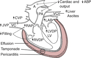
Diagnosis
Physical Examination
Laboratory Tests
Electrocardiogram
An electrocardiogram may show any of the following:
Stay updated, free articles. Join our Telegram channel

Full access? Get Clinical Tree


