Figure 11.2 The trocar is pushed through the cartilaginous rings. The novice will recognize the unexpected amount of pressure needed to penetrate the lumen of the trachea.
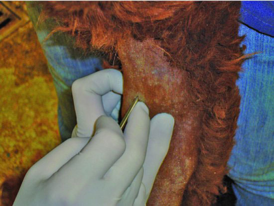
Figure 11.3 The trocar is removed, leaving the cannula sleeve. A syringe can be connected, and if no air resistance is noted when the plunger is withdrawn, correct placement has been achieved into the lumen. This author prefers to just attempt passage of the stylette, which should not meet any resistance.
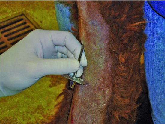
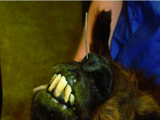
Figure 11.4 An oral speculum has been inserted between the incisors and dental pad to prevent stylette or tube damage from the molars if the animal bites down.
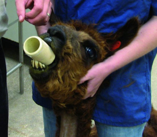
Figure 11.5 A stiff guide wire or catheter is being placed through the cannula and directed toward the oropharynx.
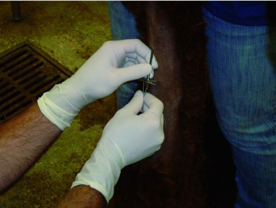
Figure 11.6 In this photograph, the guide wire has passed out the right naris. If orotracheal intubation was preferred, the wire would be withdrawn into the trachea and the head would be repositioned or a laryngoscope would be used to displace the soft palate dorsally to guide the wire orally.
Stay updated, free articles. Join our Telegram channel

Full access? Get Clinical Tree


