CHAPTER 103 Parturition and Dystocia in Swine
NORMAL PARTURITION
Maintenance of pregnancy in the sow appears to be dependent on secretion of progesterone (P4) by corpora lutea on the ovary. Estrogen levels are also increased during pregnancy (especially the last trimester) and may act to support the function of the corpus luteum (CL). Maternal plasma P4 levels are elevated throughout pregnancy and decline slowly during the last 10 to 14 days of pregnancy. The last 24 to 36 hours before parturition is marked by a more rapid decline in P4 levels. The decline in P4 concentrations coincides with the release of endogenous prostaglandin F2α (PGF2α) from the uterus. Either endogenous or exogenous PGF2α will result in the regression of a mature (greater than 12 days of age) CL. This appears to be an important endocrine signal for the initiation of parturition.1,2
Coincident with PGF2α release from the uterus is a rise in the protein hormone relaxin. This hormone is synthesized and stored within the CL of pregnancy. Relaxin levels rise from day 110 of gestation and peak 15 hours before the birth of the first piglet. Relaxin acts to soften the cervix and to inhibit myometrial contractions. Relaxin levels decline during the final moments before parturition. This decline apparently frees the myometrium to begin the contractions of labor. More PGF2α is released from the uterus during labor and apparently stimulates the release of oxytocin from the posterior pituitary of the sow. High levels of estrogen during the last month of gestation act to sensitize the myometrium to the contractile effects of oxytocin.1
Physical changes in the sow may signal impending parturition. Astute observers may notice abdominal distention as early as 80 to 90 days of gestation. Evidence of mammary development may be noted soon afterward. During the last week of gestation, the vulva may become swollen and reddened. The mammary glands become increasingly distended and may leak small amounts of colostrum during the day or two preceding farrowing. Large amounts of colostrum may be expressed from the udder within 6 to 12 hours of farrowing.3
During the 12 to 24 hours preceding farrowing, the sow becomes restless and may build a nest if bedding material is available. Alternatively, she may paw or root at the floor and bite at the bars of the farrowing crate. Other common behaviors during the last few hours before farrowing include frequent defecation and urination and an increase in respiratory rate. During the last 15 to 60 minutes before the first pig is born, physical activity will decrease and the sow will lie down in lateral recumbency. Abdominal straining may be evident and small amounts of fluid and meconium may flow from the vulva. The first piglet usually is delivered within 15 to 20 minutes.3
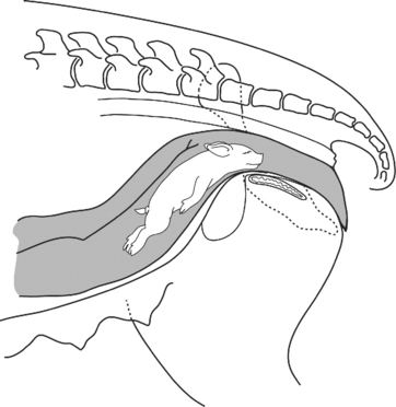
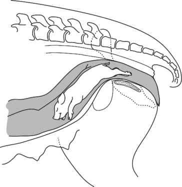
The duration of farrowing is variable but usually is complete in 1 to 5 hours. The interval between expulsion of successive piglets varies from seconds to hours but typically is 15 minutes or less. An interval of greater than 15 minutes between piglets is associated with an increased probability that the next piglet will be stillborn. Placentas may be expelled between piglets but more commonly are expelled after the birth of the last piglet in the litter. Piglets may be delivered either in a cranial presentation—head first, with front legs folded back against the body (Fig. 103-1)—or a caudal presentation—rear legs first and extended caudally (Fig. 103-2). Although both presentations are considered normal, slightly more than half of all piglets are delivered in the cranial presentation.4
DYSTOCIA
Dystocia usually is manifested by the failure to deliver piglets within an hour or 2 of the onset of labor or an interval longer than 1 hour between delivery of piglets within a litter. Other possible signs of dystocia may include prolonged gestation (beyond 116 days), illness of the sow (anorexia and depression), and discolored or fetid vulvar discharges.3
Other conditions unrelated to the bony structure of the maternal pelvis may reduce the functional diameter of the birth canal. Constipation or presence of excessive fecal material in the colon and rectum may result in partial occlusion of the birth canal. A full urinary bladder may have a similar effect. Obese sows may have fat deposits in the birth canal that decrease its diameter. Excessive manipulation and examination of the genital tract during dystocia may result in trauma and swelling of the vaginal tissues, particularly if these procedures are performed with inadequate lubrication and gentleness. A persistent remnant of the hymen also may partially occlude the birth canal.
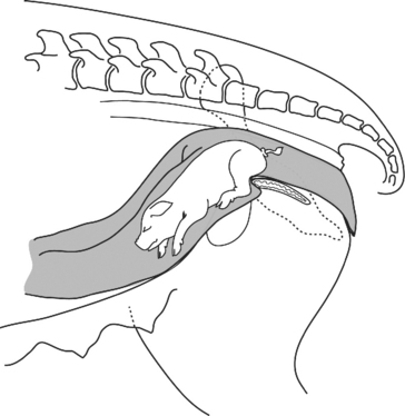
Abnormal posture or presentation of the fetus in the birth canal also may cause obstructive dystocia. In the breech presentation (Fig. 103-3), the fetus is proceeding backward or tail first through the birth canal. This differs from the normal caudal presentation in that the rear legs are flexed forward. In this position, the fetal hocks often catch on the brim of the maternal pelvis, impeding normal delivery. Occasionally, the fetus presents in a transverse or sideways position (Fig. 103-4). In this presentation the body of the fetus is flexed at the backbone or side and this part of the fetus becomes lodged in the birth canal. In the poll presentation (Fig. 103-5), the fetus comes head-first through the birth canal; however, the neck is flexed such that the top of the head (or poll) becomes lodged in the birth canal. In older sows with large litters, the uterus sometimes will be pulled down along the ventral abdominal wall and form an S-curve, with the lower part of the curve going underneath the brim of the pelvis (Fig. 103-6). A fetus may become lodged in this curve underneath the pelvis. On occasion, two fetuses may present in the birth canal at the same time (Fig. 103-7). The maternal pelvis usually is too small to allow the passage of more than one fetus at a time.3
Stay updated, free articles. Join our Telegram channel

Full access? Get Clinical Tree


