Chapter 35 Pancreatic Beta Cell Neoplasia
Tumors of the beta cells of the pancreatic islets are functional tumors that secrete excessive amounts of insulin independent of normal regulatory mechanisms. Hyperinsulinism causes hypoglycemia and corresponding clinical signs. Consider beta cell neoplasia in the differential diagnosis of hypoglycemia in middle-aged and older dogs.
ETIOLOGY
CLINICAL SIGNS
DIAGNOSIS
Signalment
Signalment is not always useful in differentiating insulinoma from other common causes of hypoglycemia (Table 35-2).
Physical Examination
The physical examination of dogs with insulin-secreting tumors is surprisingly unremarkable.
Laboratory Studies
Blood Glucose Assay
MEDICAL TREATMENT FOR ACUTE HYPOGLYCEMIC CRISIS
The acute onset of clinical signs of hypoglycemia (see Table 35-1) typically occurs in the dog in the home environment or immediately postoperatively in the dog with an inoperable tumor or metastases. Therapy depends on the severity of clinical signs and the location of the dog or cat (i.e., home or hospital).
SURGICAL TREATMENT
Surgical Anatomy
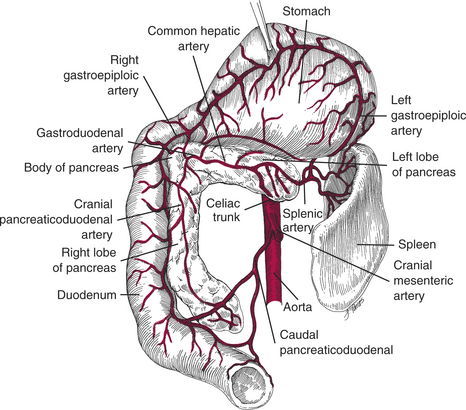
The pancreas consists of the right lobe, left lobe, and the body.
Right Lobe
The right lobe is located in the mesoduodenum adjacent to the descending duodenum. The proximal portion of the right lobe is intimately associated with the duodenum (Fig. 35-1). The proximal half of the right limb is supplied by the cranial pancreaticoduodenal artery, which is the terminal branch of the hepatic artery. The distal half of the right limb is supplied by the caudal pancreaticoduodenal artery, which is a branch of the cranial mesenteric artery. Damage to the pancreaticoduodenal vessels may result in avascular necrosis of the duodenum.
Stay updated, free articles. Join our Telegram channel

Full access? Get Clinical Tree


