36 PANCREAS
5 What hematologic changes are found in cases of acute canine pancreatitis?
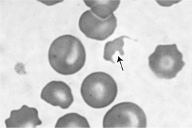
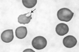
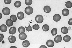
Neutrophilic leukocytosis is a common finding in cases of acute canine pancreatitis. If pancreatitis is severe (usually reflected by more severe clinical signs), the neutrophilia may be associated with a left shift and toxic neutrophils. Erythrocytosis (increased hematocrit, hemoglobin, and red blood cell [RBC] count) is common and is secondary to clinical dehydration. If a hemorrhagic pancreatitis is present, a slight anemia (nonregenerative or regenerative depending on duration of condition) may be present, but the anemia may be masked by clinical dehydration, resulting in RBC parameters within reference limits. If pancreatitis is extremely severe, disseminated intravascular coagulation (DIC) may result, possibly causing a thrombocytopenia as well as RBC pathologies on the blood smear, including schizocytes (Figure 36-1), keratocytes (Figure 36-2), and acanthocytes (Figure 36-3).

Figure 36-1 Schizocyte (arrow), a red blood cell fragment, in a dog with acute pancreatitis.
(Modified Wright’s stain; 500×.)

Figure 36-2 Keratocyte (arrow) in a dog with acute pancreatitis. Note the projections resembling blunt horns.
(Modified Wright’s stain; 500×.)
6 What changes can be expected on a routine biochemistry panel in cases of acute canine pancreatitis?
Stay updated, free articles. Join our Telegram channel

Full access? Get Clinical Tree


