Pain Assessment and Management

CHAPTER 26 Pain Assessment and Management


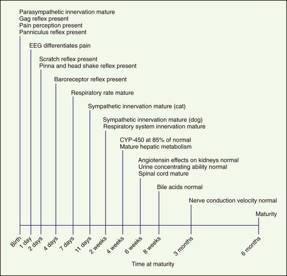
Recognition and treatment of pain in neonates is an emerging science and is controversial. To effectively treat pain in neonates, the maturity and function of many different body systems must be considered (Figure 26-1). Choosing appropriate drug therapy requires consideration of course, cause, and severity of pain and the duration and side effects of the chosen analgesic therapy (Box 26-1). In addition, other medications, concurrent medical problems and medications, and the patient’s physical status should be considered when choosing analgesics. Few clinical studies have been conducted investigating pain in neonatal dogs and cats. All too often, pain in neonates and juvenile animals is undertreated.


Physiology and Development


Development of Cardiovascular and Respiratory Systems


Parasympathetic innervation of the cardiovascular system is mature at birth. In contrast, sympathetic innervation develops over the first 2 weeks of life and reaches maturity at 14 days in dogs and 11 days in cats. The baroreceptor reflex is active as early as 4 days of age. Normal respiratory rate in dogs and cats occurs at approximately 1 week of age. During the period from birth to 1 week, the respiratory rate is increased. Innervation of the respiratory system is complete and functional by 14 days of age.


Neonates have a much larger metabolic oxygen requirement, and their carotid body chemoreceptors are immature compared with those of adult animals. Because of these factors, neonates are much more susceptible to hypoxemia. The response of a neonate to hypoxemia is a compensatory increase in tidal volume and respiratory rate. However, sleep alters this pattern by disrupting the metabolic and respiratory systems: minute ventilation and metabolic rate decrease in sleeping puppies and kittens. The result is a relative hypoxemia that is compensated for by a decrease in metabolism.


Tidal breathing in neonates is altered as a result of their increased chest wall compliance. The work of the neonate to move the diaphragm is greater and so is the need to generate increased negative pressure. As a result, respiratory depression that shortens the inspiratory phase negatively affects gas exchange and arterial blood gases (Box 26-2).


Drug Absorption, Metabolism, and Excretion


Absorption of drugs administered orally or transdermally in neonates is altered compared to adults. Gastrointestinal (GI) motility in neonates is irregular and may be related to pressure gradients in the small intestine rather than neuronal activity, until approximately 40 days of age. Generally, GI motility is decreased in the neonate, which results in decreased absorption from the stomach. However, GI permeability is also altered. Immediately after birth, the permeability of intestinal mucosa is increased, but at approximately 10 to 12 hours of age, intestinal mucosal permeability starts to decrease. The skin of neonates contains a greater percentage of water compared to adults and is also thinner, which causes an increase in absorption of drugs delivered transdermally.


Metabolism and excretion of drugs depends on liver and kidney function. Elevations and depressions in different enzyme systems associated with liver function are responsible for most differences in hepatic function in neonates. Alkaline phosphatase and gamma-glutamyltransferase (GGT) are increased in the neonate. However, gluconeogenesis, glycogenolysis, and hepatic elimination are reduced. Cytochrome P-450 activity is approximately 85% of that in an adult at 4 weeks of age and continues to increase until 8 weeks of age. Microsomal enzyme activity matures over a period of months, reaching maturity at approximately 4.5 months of age. Regardless, hepatic metabolic activity is adequate by 5 to 8 weeks of age.


Plasma protein levels in neonates are decreased compared with adults. In particular, albumin levels are lower in young animals but reach parity with adult levels by 8 weeks of age. Protein-bound drugs administered to young animals have a higher fraction of unbound and thus active drug in their plasma.


Bile flow in newborn animals is significantly decreased compared with adult animals, and bile acid composition and concentrations gradually increase. By 8 weeks of age, bile flow is comparable with an adult.


The kidneys are functionally and morphologically immature at birth and continue to develop for 2 to 3 weeks after birth. Glomerular filtration rate (GFR) and renal plasma flow (RPF) are lower in the neonate than the adult, and this correlates with a lower arterial blood pressure. As arterial blood pressure normalizes, GFR and RPF increase because GFR and RPF in neonates are directly correlated with arterial blood pressure. The effect of angiotensin on the kidneys does not reach maturity until 6 weeks of age. Urine concentrating ability also matures at approximately 6 weeks of age (Box 26-3).


Development of the Spinal Cord, Pain Receptors, and Pain Recognition


Many tactile and pain-initiated reflexes are present at birth or develop soon thereafter. The neonate’s response to noxious stimuli is appreciable from birth to 1 day of age and can be demonstrated by withdrawal from the noxious stimulus. An electroencephalogram (EEG) can differentiate this pain from other stimulation. This withdrawal from noxious stimuli in puppies is slow during the first days of life, suggesting that maturation of nociception occurs after birth. Pain sensitivity (threshold) may be more pronounced in newborns compared with adults because of an increased number of pain receptors and neural reorganization in neonates. A panniculus reflex is present at birth. The scratch reflex and the pinna and head-shake responses to noxious stimuli at the pinna or external ear canal are present at 2 days of age. The gag reflex is present at birth.


Spinal cord maturity occurs at 6 weeks of age in dogs, and the cranial portion develops more quickly than the caudal portion. Complete myelination and maturity of fasciculus gracilis and lateral corticospinal tracts occurs after 6 weeks. Maturation of the feline spinal cord and myelination of nerves occurs earlier than in dogs. Lateral columns in cats are well myelinated at 14 days of age. Dorsal nerve fibers mature rapidly during the first 8 days of life. Nerve conduction velocities in dogs reach maturity between 6 months and 1 year of age, with full maturity in cats reached in 3 months.


The initial noxious stimulus produces transduced electrical signals that are transmitted by afferent sensory neurons to projection neurons in the spinal cord, each of which has receptors that are under developmental control. In addition, these peripheral sensory neurons are overproduced during embryonic development and nearly half of these sensory neurons undergo programmed cell death in the adult. As a result, during postnatal development the perception of pain and its inhibition undergoes waxing and waning based on the neurobiologic development of the animal. This explains large interspecies and interindividual variability in the response to noxious stimuli (Box 26-4).


Physiology and Pathophysiology of Pain in Neonates


Pathophysiology of Pain


The physiology and pathophysiology of pain in neonates is similar to other advanced mammals. Peripheral nociceptors (free nerve endings of primary afferent sensory nerve fibers) transduce mechanical, chemical, and thermal energy (noxious input) into electrical impulses. There are two types of nociceptors (A-mechano-heat receptors and C mechano-heat receptors).


Nociceptive impulses are transmitted to the central nervous system (CNS) by primary afferent sensory nerve fibers. There are two main types of fibers: A-delta and C fibers. The A-delta fibers are comparably large diameter, myelinated axons that conduct impulses rapidly and are associated with “first pain.” The C fibers are smaller diameter, unmyelinated axons, with slower conduction velocities, which reinforce the immediate response of A-delta fibers. C fibers mediate second pain.


Primary sensory afferent nerve fibers synapse in the grey matter of the spinal cord dorsal horn, which is organized into layers (laminae). A-delta fibers terminate in lamina I (most important) and lamina V. The C fibers terminate in lamina II. The dorsal horn is the site of modulation of nociceptive input. The primary sensory afferent nerve fibers may form direct or indirect connections with one of three functional populations of dorsal horn neurons (interneurons, propriospinal neurons, and projection neurons). Interneurons are either excitatory or inhibitory and act as relays participating in local processing. Propriospinal neurons extend over multiple spinal segments and involve segmental reflex activity. Projection neurons participate in ascending transmission of nociceptive impulses and extend axons beyond the spinal cord to terminate in supraspinal centers (such as the midbrain or cortex). There are three types of projection neurons: nociceptive specific, wide dynamic range, and complex neurons. Communication in the dorsal horn between primary afferent fibers and dorsal horn neurons occurs by chemical signaling, mediated by excitatory or inhibitory amino acids and peptides produced, stored, and released in the nerve terminals. Glutamate and aspartate are the most important excitatory mediators. Other excitatory neurotransmitters are substance P, neurotensin, vasoactive peptides, calcitonin gene-related peptide, and cholecystokinin.


Projection neurons transmit nociceptive input to supraspinal centers via tracts that ascend in white matter of the spinal cord: spinothalamic, spinoreticular, spinomesencephalic, spinocervical, and spinohypothalamic tracts. The spinothalamic tract originates from nociceptive specific and wide dynamic range neurons in laminae I, V, VI, and VII and terminates in lateral and medial nuclei of the thalamus and is believed to be the most important with regard to nociception. The spinothalamic tract is only faintly myelinated at birth. Myelination occurs in the external tracts more quickly than in the deeper tracts, with full myelination occurring between 3 and 6 weeks of age. Myelinated fibers in grey matter laminae are present at birth. Diameter increases and myelination of these fibers continues during the neonatal period, with full myelination and density occurring at 5 weeks of age.


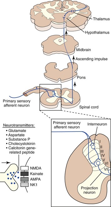
Nociceptive impulses are subjected to modulation and integration before reaching their ultimate destination, the cerebral cortex. Other supraspinal structures that process and modulate nociceptive input include the reticular formation, periaqueductal grey matter, limbic system, and the thalamus. The thalamus is the major relay station for all sensory input en route to the cerebral cortex. It is especially important for integrating nociceptive impulses and is composed of numerous complex nuclei: the lateral thalamic nuclei are involved in sensory discriminative aspects of pain, while the medial thalamic nuclei are involved in motivational affective aspects of pain. The cerebral cortex is the site where the physiologic process of nociception is integrated and ultimately perceived (Figure 26-2). Several discrete cortical regions are preferentially activated by noxious stimulation: first and second somatosensory cortices, anterior insular cortex, and anterior cingulate. Neurotransmitters in the thalamocortical region are excitatory (glutamate and aspartate) and inhibitory (gamma-aminobutyric acid [GABA], glycine, monoamines, acetylcholine, and histamine). Differences between the integration of nociceptive impulses in neonates compared with adults are unknown.


Descending inhibitory pathways modulate sensory input and are especially important for nociceptive transmission. The descending inhibitory modulatory system has four tiers, with inhibitory influences located in the following regions: cortical and thalamic structures, periaqueductal grey matter of the midbrain, rostral medulla, and spinal cord dorsal horn. Antinociceptive effects in these regions are mediated by GABA, glycine, serotonin, norepinephrine, and endogenous opioids (enkephalins, endorphins, and dynorphins). This system ensures that the physiologic pain response generated is appropriate for the noxious stimulus initiated.


Physiologic Changes and Responses to Pain


There are two important aspects to the pain experience in animals: the sensory component and the affective (the emotional) component. In addition, there is a secondary pain affect, or the consequences associated with chronic pain and homeostatic responses. Pain sensation is more intense in humans than other somatic sensations, which would seem to be an evolutionary adaptation. Additional characteristics of pain include quality (e.g., sharp or piercing, dull, throbbing), persistence, temporal summation, spatial spread of sensation at high pain levels, and unique qualities of the sensation that are intrinsically dependent on the location and degree of tissue injury.


An animal’s ability to sense noxious stimuli is present at birth, but the actual sensation of pain in animals is not assessable except by behavioral interpretation. Extrapolation of reports of pain from human studies may result in anthropomorphizing and an increase in assignment of pain in animals. Confounding this is the heightened response to what may be considered a nonpainful or mildly painful stimulus in neonates (learning). The nervous system is plastic, and extensive studies of pain in neonatal animals as a model for human neonatal pain show a heightened sensitivity to tissue injury that can result in neural reorganization by formation of new primary afferents, increased innervation of wounded tissue, and hyperexcitability of dorsal root ganglia. The long-term result of this neural reorganization is hypersensitivity to sensory input and behavioral modification.


Pain Assessment in Neonates


Assessment of pain in neonates is difficult because of an incomplete understanding of normal pain behavior in neonates. Neonatal and juvenile dogs appear to be more reactive to noxious stimuli, often demonstrating exaggerated responses compared to adult animals. This may be due to an overproduction of peripheral sensory neurons or evolutionary development of warning systems causing hypersensitivity. While the stimulus itself may be judged to be mild, the experience of the animal may be heightened as a result of innate developmental differences. Neonatal cats are less likely to demonstrate this heightened reaction, possibly the result of an earlier neurologic maturity compared to dogs.


Behavioral and physiologic parameters may be used to assess pain in neonates and are more useful in predicting stressful situations than in adults. Pain behavior in neonates and juveniles includes a change from normal behavior patterns and display of new behaviors such as positioning the painful area in such a way as to limit pressure or stimulation. In neonates, this may include a change in sleeping and eating patterns. Because this constitutes the majority of a neonate’s time, a decrease or increase in these behaviors may be indicative of pain perception. Increased vocalization (whining, whimpering, and crying) may also be an indicator of pain. Physiologic indicators of pain in neonates include changes in heart rate, respiratory rate, blood pressure, and oxygen saturation (Box 26-5).


Pain Control


Sedatives


Acepromazine


Acepromazine is a phenothiazine tranquilizer that blocks dopamine receptors in the CNS and depresses the reticular-activating system, resulting in sedation. Acepromazine also blocks alpha-adrenergic receptors. Acepromazine is not an analgesic but potentiates the effects of analgesic drugs, most notably, opioids. Acepromazine is metabolized by the liver and eliminated by the kidneys and as a result has a longer half-life in young animals. The duration of effect in adult animals is typically 4 to 8 hours and would reasonably have a longer duration of action in neonates and juveniles.


Acepromazine causes sedation without significant respiratory depression. Although systemic blood pressure can be reduced as a result of vasodilation, acepromazine administration can also result in vagally-induced bradycardia. In addition, acepromazine has antiemetic, antihistaminic, antisympathetic, antiarrhythmic, and antishock properties because of its dopamine inhibition in the chemoreceptor trigger zone.


Caution should be used when administering acepromazine to animals that are predisposed to seizures or with a seizure history because it can lower the seizure threshold. The consequences of this effect remain speculative. The dose of acepromazine should be decreased in neonates or those animals with hepatic insufficiency caused by its slower metabolism and potentially long duration. Acepromazine is a safe and effective tranquilizer in juvenile animals (Table 26-1).


TABLE 26-1 Drugs useful for treating pain in neonates


Drug Dose
Phenothiazine Tranquilizers
Acepromazine 0.01-0.05 mg/kg IM, SC
Benzodiazepines
Diazepam 0.1-0.4 mg/kg IV
Midazolam 0.1-0.4 mg/kg IV, IM
Alpha-2 Agonists
Medetomidine

Xylazine 1-2 mg/kg IM
Opiates
Morphine 0.2-1 mg/kg IM
Oxymorphone 0.02-0.2 mg/kg IV, IM, SC
Hydromorphone 0.02-0.2 mg/kg IV, IM, SC
Fentanyl 0.002-0.004 mg/kg IV, IM
Fentanyl patches


Meperidine

Stay updated, free articles. Join our Telegram channel

Sep 11, 2016 | Posted by in SMALL ANIMAL | Comments Off on Pain Assessment and Management

Full access? Get Clinical Tree

Get Clinical Tree app for offline access