Chapter 113 Orthopedic Disorders of the Distal Extremities
Disorders of the extremities distal to the carpus and tarsus usually result from direct trauma. Most abnormalities involve bone fracture and/or ligament damage associated with joint instability. The majority of fractures involve the metacarpal and metatarsal bones. Animals are usually presented with acute, non–weight-bearing lameness. Regardless of the bone being managed, internal fixation, external coaptation, or a combination of both can be used. Most surgeons prefer to use external coaptation whenever possible to manage fractures in these areas. In contrast, ligamentous injuries and associated joint instability rarely respond to external coaptation and often require surgical intervention. If fractures are open, proper open wound management is required before fracture fixation. Follow the generally accepted principles of fracture repair, including adequate reduction, alignment, and stable fixation, for the successful management of these injuries.
METACARPAL AND METATARSAL FRACTURES
Preoperative Considerations
Surgical Procedure
Technique
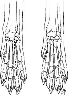
Figure 113-1 Dorsal incision lines for metacarpal fractures of a single bone (left) or multiple bones (right).
Stay updated, free articles. Join our Telegram channel

Full access? Get Clinical Tree


