Chapter 131 Ophthalmic Equipment and Techniques
OPHTHALMIC EQUIPMENT
OPHTHALMOLOGY TECHNIQUES
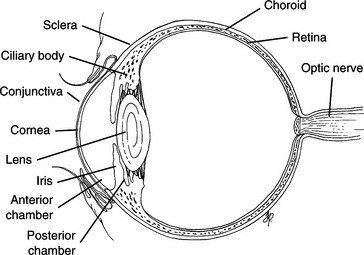
To properly evaluate and treat the patient with ocular disease, the clinician must have an accurate understanding of ophthalmic anatomy (Fig. 131-1).
Culture
Technique
1. Using a sterile swab moistened in transport media, obtain the sample in an aseptic manner from the area of concern; for example:
2. Do not use a topical anesthetic for this procedure because it may interfere with the growth of organisms.
3. Label the sample and submit for aerobic and possibly fungal culture and sensitivity testing. It should be streaked onto nutrient agar as soon as possible.
Schirmer Tear Test
Technique
2. Hold the eye closed and allow the strip to remain in place for exactly 1 minute. If convenient, both eyes may be tested at the same time.
3. Remove the strip and, using the standard measurement on the package, measure and record tear production. Normal dogs secrete 15 mm or more in 1 minute. While the normal value in the cat is reported to be similar to the dog, cats can have very low STT values without clinical signs. It is thought that stress may decrease the STT value in cats, and results must be interpreted along with clinical signs.
4. Do not use topical anesthetic for this test because the objective is to measure the response of the eye to an irritant. This requires the response of the ophthalmic branch of cranial nerve (CN) V as the afferent arm and the parasympathetic fibers in CN VII as the efferent arm.
Tear Breakup Time (TBUT)
Technique
3. Open the eyelids and, holding them open, begin timing while examining the green precorneal tear film. Continue to hold the eyelids open. The precorneal tear film should appear as a uniform, continuous film across the cornea. When the tears begin to separate into an oil and water pattern, stop timing.
Examination of the Nictitating Membrane
Technique
1. Examine the palpebral surface of the third eyelid by gently retropulsing the globe and allowing the nictitating membrane to prolapse passively while the lower eyelid is retracted. This is useful for assessing the mobility of the third eyelid and for protecting the eye when obtaining a conjunctival scraping.
2. Examine the bulbar surface of the third eyelid. This requires topical anesthesia.


Stay updated, free articles. Join our Telegram channel

Full access? Get Clinical Tree


