CHAPTER 1 History of embryology
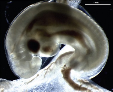
Embryology, the study of development from fertilization to birth, has always intrigued philosophers and scientists. Universal fascination with the way in which ‘life’ unfolds has led to spirited discussion of the complexities of the process amongst embryologists over the years. In 1899, Ernst Haeckel (1834–1919) considered that ‘Ontogeny is a short recapitulation of phylogeny’. In other words, ontogeny (the development of an organism from the fertilized egg to its mature form) reflects, in a matter of days or months, the origin and evolution of a species (phylogeny), a continuing process that is measured in millions of years. Although this is not entirely true, one has only to look at a 19-day-old sheep embryo to understand Haeckel’s viewpoint; the gill-like pharyngeal arches and the somites, for example (Fig. 1-1) are common to embryos of all chordates. Although trained as a physician, Haeckel abandoned his practice after reading The Origin of Species by Charles Robert Darwin (1809–1882) published in 1859 (and available at that time for fifteen shillings!). Always suspicious of teleological and mystical explanations of life, Haeckel used Darwin’s theories as ammunition for attacking entrenched religious dogma on the one hand and elaborating his own views on the other. However, in projecting phylogeny into ontogeny, Haeckel made one mistake: his mechanism of change required that formation of new characters, diagnostic of new species, occur through their addition to a basic developmental scheme. For example, because most metazoans pass through a developmental stage called a gastrula (a ball of cells with an infolding that later forms the gut) Haeckel thought that at one time an organism called a ‘gastraea’ must have existed, looking much like the gastrula stage of ontogeny. This hypothesized ancestral metazoan, he thought, gave rise to all multi-celled animals. Such a ‘single straight line’ conception of phylogeny, with all creatures standing on the shoulders of predecessors in one trajectory, has now been abandoned; phylogeny divides into a multitude of lines.
The first real embryologist that we know about was the Greek philosopher Aristotle (384–322 BC). In The Generation of Animals (ca. 350 BC) he described the different ways that animals are born: from eggs (oviparity, as in birds, frogs and most invertebrates), by live birth (viviparity, as in placental animals and some fish) or by production of an egg that hatches inside the body (ovoviviparity, which occurs in certain reptiles and sharks). It was Aristotle who also noted the two major patterns of cell division in early development: holoblastic cleavage in which the entire egg is divided into progressively smaller cells (as in frogs and mammals) and the meroblastic pattern in which only that part of the egg destined to become the embryo proper divides, with the remainder serving nutritive purposes (as in birds). The fetal membranes and the umbilical cord in cattle were described by Aristotle and he recognized their importance for fetal nutrition. By sequential studies of fertilized chick eggs, Aristotle made the very important observation that the embryo develops its organ systems gradually – they are not preformed. This concept of de novo formation of embryonic structures, which is referred to as epigenesis, remained enormously controversial for more than 2000 years before becoming fully accepted; Aristotle was way ahead of his time. The enigma of sexual reproduction also intrigued Aristotle. He realized that both sexes are needed for conception but felt that the male’s semen did not contribute to conception physically, but by providing an unknown form-giving force that interacts with menstrual blood in the womb of the female to materialize as an embryo. In this case Aristotle was wrong. Ironically, in contrast to the non-acceptance of his correct views on epigenesis, his error about conception prevailed for about 2000 years, and it took a battle of almost a hundred years to correct it!
During those 2000 years, the science of embryology developed very slowly along descriptive anatomical lines. One of the first anatomical descriptions of the pregnant uterus of the pig was by Kopho (years of birth and death unknown) in the early 13th century in his work Anatomio Porci. Kopho worked at the famous medical school of Salerno using pigs as models because human cadavers could not be dissected for religious reasons. During the early Renaissance, the famous and incomparably artistic anatomical studies of Leonardo da Vinci (1452–1519) included investigation of the pregnant uterus of a cow. His drawings of a pregnant bicornuate uterus and of the fetus and fetal membranes released from the uterus are reproduced in Fig. 1-2. In an accompanying drawing, Leonardo depicted a human uterus cut open to ‘reveal’, not a human placenta but a multiplex, villous, ruminant placenta (see Chapter 9)! Clearly it had not been possible for him to actually study a human pregnancy. The structures are described in the characteristic mirrored writing of Leonardo, who worked mostly in Florence.
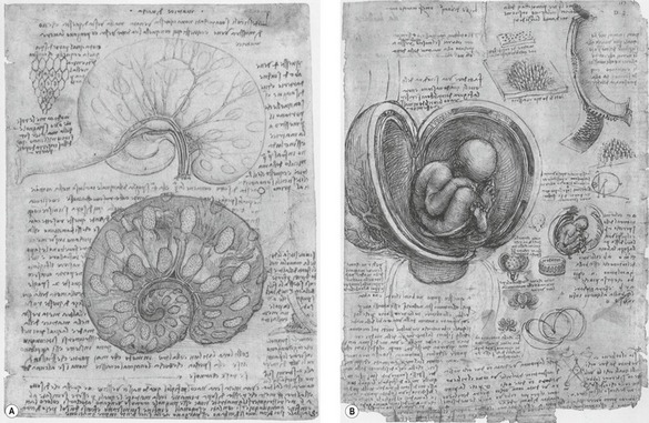
Fig. 1-2: Drawings of Leonardo da Vinci. A: Top: A pregnant bicornuate uterus of the cow. Bottom: Fetus and fetal membranes showing the cotyledons of the placenta (see Chapter 9). B: Opened simplex uterus of human showing the fetus with the umbilical cord. Note that the placenta is of the ruminant multiplex type with several placentomes drawn on the cut wall of the uterus and, at the right top, a single cotyledon drawn at a higher magnification displaying its villous surface.
The Renaissance, from the 13th to the 15th century, was a great time for anatomical studies and, happily, coincided with the invention of book printing by Johann Gutenberg. Thus, the first major publication on comparative embryology was De Formato Foetu in 1600 by the Italian anatomist Hieronymus Fabricius of Acquapendente (1533–1619). Fabricius described and illustrated the gross anatomy of embryos and their membranes in that book, but was not actually the first to do so; another Italian anatomist, Bartolomeo Eustachius (1514–1574), had previously published illustrations of dog and sheep embryos in 1552. We now recognize the names of Fabricius, in the term bursa Fabricii (the immunologically competent portion of the bird gut), and of Eustachius in the Eustachian tube.
One of the earliest and most influential names in the fascinating story of the discovery of the mammalian egg was that of William Harvey (1578–1657), personal physician to the English kings James I and Charles I, and famous for his description of the circulation of the blood. In 1651, Harvey published De Generatione Animalium (Disputations touching the Generation of Animals) with a famous frontispiece showing Zeus freeing all creation from an egg bearing the inscription Ex ovo omnia (All things come from the egg). However, it should be realized that, far from advancing 17th century knowledge of reproduction and embryology, Harvey’s observations in some ways impeded progress. From having studied with Fabricius, Harvey was imbued with Aristotle’s view that the semen provided a force that interacted with the menstrual blood to materialize as an embryo. Harvey set out to understand this process by looking for the earliest products of conception in female deer killed during the breeding season in the course of King Charles I’s hunts in his Royal forests and parks over a 12-year period. In the red and fallow deer that he studied, the male’s rut begins in mid-September and so Harvey dissected uteri throughout the months of September to December. Believing, wrongly, that copulation coincides with the onset of the rut, Harvey was mystified to find nothing that he recognized as an embryo until mid-November, some two months later. This forced him to the erroneous, but entirely logical conclusion that ‘nothing after coition is to be found in [the] uterus for many days together’. When he did find a conceptus, that, for Harvey, was the egg: ‘Aristotle’s definition of an egg applies to it, namely, an egg is that out of a part of which an animal is begotten and the remainder is the food for that which is begotten’.
As it was, the first scientist to actually see the mammalian egg (which everyone believed to exist, but no one had seen) was the Estonian medical doctor Karl Ernst von Baer (1792–1876). He opened the ‘Graafian egg’, as the follicle was known at that time, and saw with his naked eye a small yellow point which he released and examined under the microscope (Baer, 1827). There, upon a first glance, Baer was stunned and could hardly believe that he had found what so many famous scientists including Harvey, de Graaf, Purkinje and others had failed to find. He was so overcome that he had to work up courage to look into the microscope a second time. The mammalian egg had been identified.
How about the spermatozoa? Anton van Leeuwenhoek (1632–1723), a Dutch tradesman and scientist from Delft, was the first to report having seen moving spermatozoa. He constructed a single lens microscope that magnified up to about 300 times. Technically, this microscope was an amazing achievement – no bigger than a small postage stamp and resembling a primitive micromanipulator. It was used close to the eye like a magnifying glass. Using this, Leeuwenhoek drew spermatozoa from different species. Initially, Leeuwenhoek was reluctant to study sperm and he questioned the propriety of writing about semen and intercourse. When he first focused his microscope on semen, Leeuwenhoek discovered what he then took to be globules. However, he so disliked the prospect of having to discuss his findings that he quickly turned to other matters. Three or four years later, however, in 1677, a student from the medical school at Leiden brought him a specimen of semen in which he had found small animals with tails, which Leeuwenhoek now observed as well. Consequently, Leeuwenhoek resumed his own observations and, in his own semen (acquired, he stressed, not by sinfully defiling himself but as a natural consequence of conjugal coitus), observed a multitude of ‘animalcules,’ less than a millionth the size of a coarse grain of sand and with thin, undulating, transparent tails. A month later, Leeuwenhoek described these observations in a brief letter to Lord Brouncker, president of the Royal Society in London. Still uneasy about the subject matter, he begged Brouncker not to publish it if he thought it would give offence. Leeuwenhoek’s observations prompted vivid discussions and controversies on the significance of these living objects. At first, it was commonly held that the tadpole-like creatures were parasites. Leeuwenhoek may have been biased towards that view as he was actually engaged in parallel studies of parasites and was the first to see Giardia, a protozoan parasite that infects the gastrointestinal tract. Giardia are flagellated and may, at poor microscopical resolution, resemble spermatozoa. Other scientists considered that the whirling action of the objects was meant to prevent solidification of the semen. In the preformation school, however, Leeuwenhoek’s observations encouraged the thought that the sperm head contained preformed miniatures of babies, foals, calves etc. and so these scientists became known as the spermists.
In spite of Wolff’s contribution, the preformation theory persisted until the 1820s when new techniques for tissue staining and microscopy allowed a further advance in the science of embryology. Three friends, Christian Pander (1794–1865), Karl Ernst von Baer, and Martin Heinrich Rathke (1793–1860), all of whom came from the Baltic region and studied in Germany, formulated concepts of great relevance for contemporary embryology. Pander expanded the observations made by Wolff and also, despite studying the chick embryo for only some 15 months before becoming a palaeontologist, discovered the germ layers (Pander, 1817). The overall term ‘germ layers’ is derived from the Latin germen (‘bud’ or ‘sprout’) whereas the three individual layers are of Greek origin: ectoderm from ectos (‘outside’) and derma (’skin’), mesoderm from mesos (‘middle’), and endoderm from endon (‘within’). Pander also noted that organs were not formed from a single germ layer. A remarkable feature of Pander’s book from 1817 is the quality of the illustrations drawn by the German anatomist and artist Eduard Joseph d’Alton (1772–1840); they beautifully depict details that had not yet been defined (Fig. 1-3). This classical work underlines the necessity for precise observational skills in embryology.
< div class='tao-gold-member'>