Nuclear Scintigraphy
Basic Information 
Overview and Goal(s)
• Medical nuclear medicine is the science of using radioactive substances (termed radionuclides or radioactive isotopes) to evaluate specific metabolism and physiology. As a radionuclide decomposes (to a more stable state) it releases energy in the form of radioactivity, termed radioactive decay. This emitted energy can be captured and quantified in assessment of various disease processes.
• The most commonly performed nuclear medicine study in equine practice is a bone scan. Bone scans are useful for identifying occult or nonlocalizable causes of lameness referable to the musculoskeletal system.
• Bone scans are performed by injecting bone-seeking radiopharmaceuticals that allow detection of abnormal bone metabolism and/or altered blood flow. The most commonly used radiopharmaceutical is technetium-99m (99mTc, commonly referred to as “tech”). During the decay process, 140keV gamma rays are emitted, easily detected for diagnostic imaging purposes using a gamma camera. Gamma rays are identical to x-rays, differing only in origin (nuclear vs. x-ray machine, respectively).
• Radiopharmaceuticals are drugs (compounds or materials) labeled (“tagged” or linked) to a radionuclide. In many cases, radiopharmaceuticals function much like materials found in the body and do not produce special pharmacologic effects.
• For bone scans, 99mTc is labeled to an inorganic phosphate that is essential for bone metabolism. The most commonly used radiopharmaceutical for bone scans is 99mTc methylene diphosphonate (99mTc-MDP); 99mTc hydroxymethylene diphosphonate (99mTc-HMDP) is less commonly used.
• There are three phases of a musculoskeletal nuclear medicine scan, classified by the temporal sequence of the scan: vascular, soft tissue, and bone phases.
 In the vascular phase, the region of interest is scanned immediately after injection of the radiopharmaceutical. As the name implies, this phase assesses arterial and venous blood flow. It lasts 1 to 2 minutes and is rapidly replaced by the soft tissue phase. The vascular phase is not commonly performed.

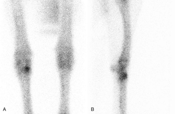
 Incorporation of radiopharmaceutical occurs in all areas of living bone, but more so in bone that is actively remodeling. As the radiopharmaceutical decays, more gamma ray emission is detected in areas of increased bone turnover, thereby allowing localization of active remodeling. This is termed a hot spot (Figure 1). Occasionally pathologic decreased or absent bone metabolism is present, termed a cold spot (eg, a bone infarction).
• Nuclear imaging studies can yield valuable information regarding the site of abnormal blood flow and altered physiology, but the images have relatively poor anatomic detail.
• Rarely can a specific diagnosis be made based on abnormal radiopharmaceutical uptake identified on a bone scan; however, these studies can direct the clinician to one or a few limited anatomic regions for further imaging and clinical evaluation.
Equipment, Anesthesia
• The patient should be standing squarely for the examination.
• Sedation is necessary to avoid motion artifact.
• The distance between the patient and the gamma camera surface is important and should be minimized at all times.
 Increased patient distance from the gamma camera leads to a decrease in detectable radiopharmaceutical uptake, degrading image quality.
 When imaging bilateral structures simultaneously (Figure 2; also see Figure 1, A) it is important that the structures are equidistant from the gamma camera to avoid differences in apparent radiopharmaceutical uptake.
• A lead shield should be used to block gamma radiation from reaching the gamma camera from nonimaged contralateral limbs. For example, a lead blocker should be used along the medial aspect of the left front leg when obtaining a lateral image of the right front leg (see Figure 1, B).
Stay updated, free articles. Join our Telegram channel

Full access? Get Clinical Tree


