Chapter 99 Neoplasia of the Maxilla and Mandible
ETIOLOGY
The oropharyngeal region is the fourth most common site of malignant neoplasia in the dog.
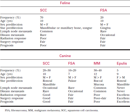
Morbidity and mortality often result from local disease rather than from distant metastasis. Therefore, local tumor control is paramount to a successful outcome. Surgical resection should be considered a first line of treatment for all oral neoplasms. See Table 99-1 for a summary of oral neoplasia in dogs and cats.
DIAGNOSIS
History
• Most patients with oral neoplasms present with a chief complaint of drooling, halitosis, and dysphagia.
• Oral bleeding may occasionally be seen, and some owners report seeing blood in the water dish after the pet drinks.
Physical Examination
• If the tumor is associated with the temporomandibular joint (TMJ), vertical ramus of the mandible, or caudal pharyngeal region, pain may be elicited when trying to open the mouth.
• Many patients do not allow a complete oral examination while they are awake due to pain; consider sedation of patients.
Diagnostic Evaluation
Imaging Studies
• Obtain skull radiographs under anesthesia to evaluate the extent of bony involvement into the maxilla or mandible. A skull series (lateral, ventral-dorsal, right and left oblique views) is required to fully evaluate the skull (see Chapter 4). Radiographic abnormalities include changes in cortical bone density, periosteal new bone formation, involvement of adjacent soft tissues, and loose teeth.
• Computed tomography (CT) scanning and magnetic resonance imaging (MRI) of the skull is very helpful in evaluating extent of tumor invasion and presurgical planning. These imaging modalities are especially helpful in evaluating caudal maxillary tumors or tumors involving the orbit, zygoma, TMJ, or vertical ramus of the mandible.
Biopsy
• Biopsy is very important in determining the treatment, as well as the long-term prognosis, for the pet. Many oral tumors do not exfoliate easily, so fine-needle aspiration is usually not helpful. A wedge biopsy for histopathologic examination is necessary for a definitive diagnosis.
• Obtain fine-needle aspirates for cytology or histopathologic evaluation via biopsy of all enlarged, submandibular, or pharyngeal lymph nodes to look for metastatic disease. Perform fine-needle aspiration even on normal-sized regional lymph nodes. In one study, 40% of dogs with oral melanoma had normal-sized lymph nodes and had evidence of metastasis to the lymph nodes on cytologic evaluation.
TUMOR TYPES
Benign Non-odontogenic Neoplasms
Epulis
Three types of epulides are characterized, based on histopathologic evaluation:
• Fibromatous epulis is a benign, noninvasive growth in which the periodontal ligament stroma is the predominant cell type. These epulides are pedunculated and often multiple.
• Ossifying epulides have a similar biologic behavior to fibromatous epulides, but histopathologically they have an osteoid component. Malignant transformation to osteosarcoma has been reported.
• Acanthomatous epulis is the most aggressive of the epulides. This type of epulis is characterized by extensive bony invasion into the alveolar bone and is most often seen in the rostral mandible. Acanthomatous epulides can become quite large but do not metastasize. However, approximately 30% of acanthomatous epulides undergo malignant transformation.
Treatment and Prognosis
• Since epulides are local tumors, they can be treated with aggressive curettage of the alveolar socket (with fibromatous and ossifying epulides). However, a better outcome can be achieved with en bloc resection of the tumor and surrounding bone. En bloc resection is recommended with acanthomatous epulides due to their extensive bony involvement. Recurrence rate is high if the tumor is only debulked, and a complete resection is considered curative.
Malignant Non-odontogenic Neoplasms
Malignant Melanoma
Treatment and Prognosis
• Treatment of choice is early, complete en bloc surgical resection. Tumor size is important, as melanomas less than 50 mm2 are associated with a better outcome.
• High-dose, fractionated radiation therapy has shown moderate success in treatment. One study showed an increase in survival time with surgery and radiation therapy when compared with surgery alone.
Squamous Cell Carcinoma
Fibrosarcoma
• Fibrosarcoma is the third most common tumor behind malignant melanoma and SCC, and it is the second most common tumor in cats.
• Fibrosarcomas are firm, slow-growing tumors that are characterized by locally aggressive behavior and late metastasis.
• Fibrosarcomas are seen most commonly on the gingival margin of the maxilla, between the upper canine tooth and the fourth premolar.
• Due to extensive tissue invasion, local recurrence is common, especially in tumors that cross the midline of the hard palate or are caudally located.


