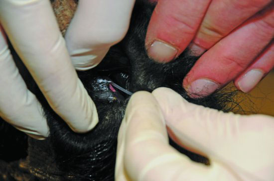
Figure 68.2 The passage of a 5-French polypropylene catheter in a retrograde fashion into the nasal punctum to facilitate flushing of the NL duct.

For animals with suspected atresia or unresolved occlusion of the duct, contrast media may be infused through the catheter and radiographs obtained to determine the extent of the blockage or atresia. In most cria with congenital nasolacrimal duct atresia, the nasal puncta is not present. In these cases, a 3.5-French polypropylene catheter is passed via the inferior conjunctival puncta, and saline is pulsed while performing an intranasal examination. In most cases of atresia, a membrane can be seen pulsing over the site of the nasal puncta. In these cases, a #15 scalpel blade or 14 gauge needle can be used to puncture the membrane. Then, a saline flush is continued until the duct is thoroughly cleared and the catheter withdrawn. In atresia cases where no nasal puncta can be observed, surgical conjunctivorhinostomy may be required to establish tear flow.
Stay updated, free articles. Join our Telegram channel

Full access? Get Clinical Tree


