8 MORPHOLOGY, FUNCTION, AND KINETICS
1 Describe the morphology of neutrophils from animals.
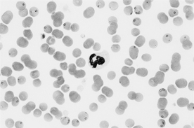
Neutrophils are the granulocytic leukocytes with specific granules that are usually inconspicuous or “neutral” in color. The specific granules of neutrophils from primates (including humans) stain more prominently, whereas in bovine neutrophils the cytoplasmic color is pink because of large tertiary granules. The neutrophil nucleus has darkly condensed chromatin and is segmented into three to five lobes interspersed by nuclear constrictions. In horses the individual lobes are further lobulated, producing a jagged or more segmented nuclear profile. On blood smears, neutrophils appear slightly smaller than the other granulocytic leukocytes, eosinophils and basophils (Figure 8-1).
2 What are heterophils?
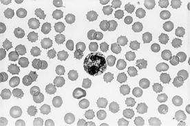
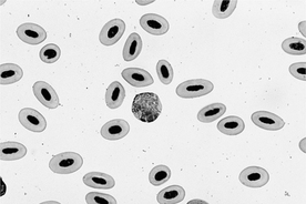
In some small mammals the specific granules in neutrophils stain eosinophilic, so their neutrophils are termed heterophils. In rabbits the granules in heterophils are very prominent and, at first glance of a blood smear, more closely resemble eosinophils (Figure 8-2). The eosinophils in rabbits are distinguished by their more intensely eosinophilic granules. In guinea pigs and hamsters the granules in heterophils are less prominent. In birds and reptiles the leukocytes considered the counterpart of neutrophils are also called heterophils. The heterophils of birds and reptiles contain large, prominent eosinophilic granules that are fusiform or round (Figure 8-3).
3 What is the “drumstick appendage”?
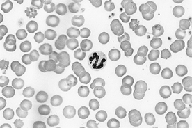
The drumstick appendage is a small, round nuclear lobe connected to the neutrophil nucleus by a thin filament (Figure 8-4). It is observed normally in a low percentage of the neutrophils in females and represents the inactivated second X chromosome. This structure corresponds to the Barr body found in epithelial cells of females. If observed in a significant number of the neutrophils in a male, the drumstick appendage suggests a chromosomal disorder, such as the XXY syndrome in male calico cats.
5 Describe the morphology of eosinophils.
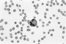
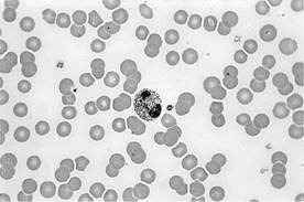
Eosinophils contain prominent specific granules in the cytoplasm that stain eosinophilic. The nucleus is lobulated as in other granulocytic leukocytes but is typically less lobulated than in neutrophils. The appearance of the cytoplasmic granules in eosinophils is unique for the domestic animals (Figures 8-5 and 8-6): rod shaped in cats, uniformly small and round in ruminants and pigs, very large and round in horses, and round but variable in number and size in dogs. Some canine eosinophils may contain as few as one or two very large granules. In contrast, the eosinophils of greyhounds and some other dogs lack eosinophilic granules, with clear vacuoles in the lightly basophilic cytoplasm.
8 Describe the morphology of basophils.
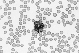
The specific granules in basophils typically stain metachromatic (meaning “other color”), the unique color produced by hematologic stains that is purple rather than basophilic. The metachromatic staining characteristic of basophils is attributed to the proteoglycan content of their granules. The metachromatic granules in bovine, equine, and porcine basophils are numerous and can obscure the segmented nucleus (Figure 8-7). There are relatively low numbers of cytoplasmic granules in dogs, so the diffuse basophilia of the cytoplasm is more apparent. Feline basophils are unusual because the specific granules lose their metachromatic staining in basophil precursors and appear lavender or gray in the mature basophil (Figure 8-8).
Stay updated, free articles. Join our Telegram channel

Full access? Get Clinical Tree


