Fig. 1.
H&E staining of a coronal slice of rat brain 1 month after injection of whole blood. *Hematoma cavity.
3.5.2 Hippocampus Swelling and Atrophy (Fig. 2)
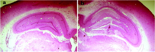
Fig. 2.
H&E staining showing the ipsilateral and contralateral rat hippocampus 24 h after intrahippocampal injection of iron.
Measure bilateral hippocampus three times and get an average size (4).
Expressed as a percentage of the ipsilateral/contralateral of swelling or atrophy.
3.5.3 Pig White Matter and Hemisphere Measurement (Fig. 3)
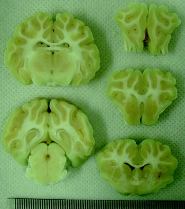
Fig. 3.
Pig coronal brain sections 2 months after sham operation.
Measure the bilateral white matter and hemisphere and multiplying by the thickness (8 mm) of the sections.
Measure three times and get a mean value.< div class='tao-gold-member'>Only gold members can continue reading. Log In or Register to continue
Stay updated, free articles. Join our Telegram channel

Full access? Get Clinical Tree


