3 Microbiology
You cannot. Bacteria are everywhere, and every time a dog breathes, sits, eats, or moves, it is exposed to bacteria. To treat all the dogs in a kennel with antibiotics in an attempt to eradicate all bacteria is foolhardy because all the dogs will be reinfected as soon as the antibiotic is out of their systems.
Mycoplasmata are part of the normal bacteria in the respiratory and reproductive tracts of dogs. They can cause reproductive tract disease in dogs, but you are as likely to culture Mycoplasma organisms from a normal dog as you are to culture them from a dog with a reproductive problem. I believe that the majority of dogs from which Mycoplasma organisms are cultured do not have disease resulting from the organisms, and to treat that dog, much less the whole kennel, is foolhardy.
Vaginal cultures may be useful in helping diagnose low-level uterine infection. We cannot access the uterus, but if we collect a specimen of the bloody discharge of heat from the vagina during proestrus, we can get an idea of what’s washing down out of the uterus. I do not do vaginal cultures routinely but always perform them on bitches with a history of a reproductive problem, such as pregnancy loss, infertility, or uterine disease. I am not a proponent of automatically treating bitches with antibiotics prebreeding and much prefer a culture sample be drawn first to guide the choice of antibiotic used, if any.
During natural service, bacteria are transferred from the bitch to the dog and vice versa. However, the normal bacteria in the vagina and on the surface of the penis are in a balance, and “intruders” quickly are overgrown and removed. For virtually all disease of the reproductive tract associated with bacterial infection, it is the animal’s own bacteria that cause the infection, with one organism upsetting the balance by growing to an abnormally large extent.
Microbiology is the study of microorganisms, free-living life forms that may exist as a single cell or a cell cluster. Some microorganisms can reproduce independently, whereas others require a multicellular organism as a host.
Microorganisms are on every surface and live in many areas of the body. In the environment, microorganisms break down complex molecules into smaller products or nutrients, some of which are vital for plant growth. Some of the compounds produced by microorganisms are useful as drugs. For example, the antibiotic streptomycin is produced by bacteria of the genus Streptomyces, and penicillin is produced by the fungus Penicillium. There are no environments on earth in which no microorganisms exist and some in which only microorganisms exist, such as the arctic and deep in the ocean.
Control of undesirable microorganisms in the environment is accomplished with the use of sterilants or disinfectants. Sterilization is destruction of all microorganisms. Disinfection is destruction or inactivation of potentially infective organisms. Examples of sterilants and disinfectants include the following:
Microorganisms in the body metabolize nutrients. The presence of the normal bacterial population in a given area of the body prevents growth of abnormal bacteria that presumably could cause harm. Areas of the body that always harbor some bacteria are the skin, nasal cavity, mouth and throat, vagina, end of the urethra, penile and preputial mucosa, and intestinal tract. Control of microorganisms in the body requires use of antibiotics (see Chapter 2).
I. TYPES OF ORGANISMS
A. BACTERIA
Bacteria are free-living organisms that can reproduce wherever conditions are appropriate. A given bacterial species requires specific temperature, pressure, moisture, and air conditions to survive. Organisms of interest to veterinary medicine generally do not thrive in very hot or very dry conditions and are easily destroyed by common antiseptic agents, like bleach.
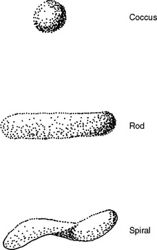
Bacteria can be classified by shape, by atmosphere in which they grow, or by staining characteristics. Common bacterial shapes are rods, cocci, and spirals (Figure 3-1). Aerobic bacteria are those that require air to reproduce; anaerobic bacteria are those that do not reproduce in air.
Gram staining is a differential staining with safranin, which stains some bacteria pink, and crystal violet, which stains some bacteria purple. A specimen is smeared on a glass slide and dried and fixed with heat. The slide is stained with crystal violet, then washed and treated with iodine. Then the slide is decolorized with alcohol or acetone and the slide stained with safranin. Those bacteria classified as gram-positive (gram+) do not decolorize and so retain the purple color; those classified as gram-negative (gram-) stain pink with safranin.
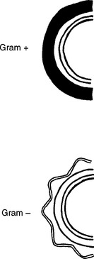
Bacteria show this differential staining because of differences in their cell wall. All bacteria have a rigid cell wall composed of compounds called peptidoglycans and lipopolysaccharides. These compounds control movement of water and other molecules in and out of the bacterial cell, protecting the nucleus and organelles within. The nucleus contains the genetic material of the cell, and the organelles generate energy, synthesize protein, and metabolize nutrients within the cell. Gram+ bacteria have a thick peptidoglycan layer. Gram- bacteria have a thin peptidoglycan layer and thick lipopolysaccharide layer (Figure 3-2).
Bacteria can be identified by culture. It is important to remember that some areas of the body have a normal bacterial population. In general, bacterial growth from those areas is considered significant only if there is heavy growth of a single organism. A good example is vaginal culture of the bitch. Positive vaginal cultures mean nothing because there is a normal bacterial population in the vagina. Vaginal cultures may be a reflection of uterine disease only in the following situations:

Stay updated, free articles. Join our Telegram channel

Full access? Get Clinical Tree

