MARSUPIALS

CHAPTER 11 MARSUPIALS


HUSBANDRY


Tammar Wallabies


Because tammar wallabies (Macropus eugenii) are small macropods, this species can be maintained within large groups in relatively small enclosures. As these animals browse heavily on grass, it is important that they have yards for periodic grazing. Tammar wallabies will eat bark they can access at the base of trees; therefore, tree guards made of wire are advisable. Pelleted food similar to what might be fed to domestic ruminants is often the primary diet component for these animals in captive settings. As stated, grass is advisable as a supplemental feed, but if grass is not available, alfalfa hay can be provided as an alternative.


The natural habitat of the tammar wallaby is low-growing and coastal scrub, eucalyptus species, woodland thickets, and sclerophyll forest. As such, it is important to provide shelter and places to hide, using branches and large, hollow concrete tubing in their yards.


The smaller size of the tammar wallaby makes it easier to catch than its larger counterparts. As with all exotic species, tammar wallaby captures should be conducted by experienced personnel and planned well in advance to minimize stress to the animal(s). An efficient capture will minimize the chance of injury that might otherwise occur from running into fences or other animals within the enclosure. A long-handled net can be used for the initial capture. Removal of the animal from the net is achieved by holding the base of the tail and holding the animal on its back. Animals can then be placed into a closed mesh bag. The bag should be as lightweight as possible to avoid any undue weight on the animal. Once restrained in the relative darkness of the bag, the animal will usually become calm, allowing for easier examination.


One of the most common reasons for examining these animals in captivity is to monitor breeding and pregnancy. Examination should be carried out while the animal is in a Hessian bag, again holding at the base of the tail and placing the animal on its back. The back legs should be free of the bag to prevent injury and allow better exposure of the pouch. The appearance of the pouch can be an indicator of the reproductive state of the animal. The pouch normally has a “dirty” appearance and is covered in a dark, tacky secretion. If the pouch appears moist and clean, then the female has likely been licking it in anticipation of an imminent birth. The presence of an elongated teat in an empty pouch indicates ongoing suckling of an “on foot” pouch young. Female tammars are capable of nursing two pouch young at the same time, one newborn and one on foot.


Fat-Tailed Dunnart


Fat-tailed dunnarts (Sminthopsis crassicaudita) can be maintained in windowless rooms with artificial light and at a constant temperature of 20° C to 24° C. Multiple rooms can be designated, each with a particular purpose. One room can have a reduced day length of 8 hours in preparation for stimulating males to breed. Other rooms to include are a nursery/maintain room and a breeding room—each with a 16-hour-day length. Galvanized metal breeding cages (∼50 ×35 ×20 cm) with hinged mesh tops, removable plate glass fronts, and metal base trays are advisable for this species. Nest boxes should be plastic (for ease of cleaning) and well ventilated, with shredded paper as a substrate. Animals that are able to eat on their own can be housed in standard laboratory rat cages. Plastic mouse wheels should be provided for breeding animals but not for nursing animals. It is essential to provide dry, clean autoclaved sand in cage trays for animals to groom themselves.


The natural diet of the fat-tailed dunnart is mainly comprised of invertebrate species (e.g., locusts, moths, cockroaches, centipedes, and scorpions) and vertebrates (e.g., small lizards and baby mice). In the laboratory setting, the dunnart can be fed canned pet food, dry cat food, and mealworms. Water and dry cat food should be fed ad libitum.


Restraint of the dunnart requires holding the tail and supporting the body. Brief restraint is accomplished by grasping the scruff of the neck. Pouch and health checks should not be conducted more than once a week to limit stress.


Brushtail Opossum


It is the habit of the brushtail opossum (Trichosurus vulpecula) to remain solitary, with the exceptions of the mating period and when nesting and feeding sites are limited. Cage styles include individual wire cages (1 m ×0.4 m ×0.4 m or 1 m ×0.4 m ×0.55 m) with mesh floors and a removable nest box (0.35 m ×0.2 m ×0.2 m), galvanized wire rabbit cages (0.5 m ×0.3 m ×0.55 m), and stainless steel wire cages (0.76 m ×0.6 m ×0.6 m). To ensure seclusion, solid sides can be provided with a drape over the cage front. Catch trays under cages should be cleaned and waste food removed once daily. Nest boxes should be cleaned at least once a week.


Group housing is possible for the brushtail opossum. Wire mesh pens with grass or concrete floors work well. Outdoor enclosures should be well built and impenetrable to birds or vermin. Enclosure sizes for breeding are approximately 11 m2 for two animals, 16 m2 for three to four animals, and 8 m2 for up to seven animals. Identification of group-housed animals can be a challenge, and therefore individual animals should be identified by a small numbered metal ear tag or some other means of permanent identification. Opossums can maintain a body temperature of 36° C to 37° C in ambient temperatures from 10° C to 30° C. Yet, it is important that outdoor pens have shelters for the opossums to escape wind and rain. Nesting boxes or Hessian nesting sacks should be provided for each individual. Perches and branches will provide enrichment and reduce aggression. In particularly warm climates, ventilation and insulation may be required.


Opossums are hindgut fermenters, and as such, their digestive system is best equipped to handle a high-fiber diet. The cecum and proximal colon are well developed and serve as the primary sites of microbial fermentation. The brushtail opossum is predominantly an herbivore. In the wild, leaves, blossoms, and fruit from a wide range of tree species are consumed. Grass, clover, and broadleaf weeds also are part of their natural diet. They have been known to eat small birds, eggs, and invertebrates as well. In the captive situation, a variety of fruits and vegetables, eucalyptus, and other leafy vegetation can be offered. Rabbit or stock feed pellets may be offered in a waterproof hopper, and water in a self-filling trough is appropriate for ad libitum administration in the outdoor environment. Opossums are susceptible to calcium toxicity and subsequent calcinosis; therefore, food with high calcium content should be avoided. Brushtails tend to be wasteful feeders, but on average, a 3.0 kg animal eats approximately 60 to 160 g of pellets daily. Adult live weights range between 1400 and 1600 g depending on diet, exercise, and genetic background.


Sugar Gliders


Sugar gliders (Petaurus breviceps) are best maintained in groups of two or more. Cage size should be appropriate to the number of animals being housed within the cage. For example, a 50 ×50 ×75-cm cage would accommodate two sugar gliders, and a 2 ×2 ×2-m aviary-type cage would hold up to six sugar gliders. Wire openings should be no greater than 2 ×2 cm. If no horizontal bars are present, vertical bars should be spaced no more than 6 mm apart. Galvanized wire should be avoided, as galvanization incorporates zinc and lead components, and therefore the potential for toxicity exists if these elements are consumed over time. Enrichment and use of cage space can be encouraged by providing tree branches and an exercise wheel 25 cm in diameter. It is important that the wheel is solid and not wire to avoid tail entrapment. In the wild, sugar gliders will hide in leafy nests found in the hollows of trees. This can be recreated in captivity by providing a hollow log or small wooden box. A 25 ×10 ×15-cm nest box with a 5-cm diameter entrance hole is appropriate for about six sugar gliders. The size of the nest box can be a determinant of group size in captivity. The nest box should be cleaned on a regular basis. A hinged top allows easy accessibility for capture and examination of the captive glider. It is important to note that sugar gliders will chew wooden structures, so nest boxes made of wood may need to be replaced.


A piece of cloth or sock placed in a nest box will provide comfort and security to a sugar glider. Owners may make the common error of using cedar or pine shavings. These wood products should be avoided because the aromatic oils in these products can prove to be toxic.


On average, owners will keep sugar gliders at temperatures between 64° F and 75° F (18° C to 24° C). However, this is the lower end of the normal ambient temperature for these animals; supplemental heat for these animals must be provided. Caution should be taken to prevent thermal burns that may occur with an additional heat source. Sugar gliders can withstand temperatures up to 88° F (31° C), but any warmer and hyperthermia is likely to result.


PERFORMING A PHYSICAL EXAMINATION


The diversity of marsupial species presented to the veterinary practitioner appears to be increasing. Before initiating a clinical examination on any marsupial species, it is the responsibility of the practitioner to be familiar with the normal appearance and behavior of the species he or she is examining.


The sugar glider is gaining popularity as a companion animal and is one of the marsupials being presented more frequently to veterinary practice. In the wild, the sugar glider is known to inhabit the forests of Australia, New Guinea, and Tasmania. Its body averages 7 inches in length, with another 8 inches of tail.


The sugar glider has a thick, gray coat with a distinct black stripe running down its spine. Its ability to glide from tree to tree is assisted by a membrane of skin known as a patagium, which connects the lateral aspect of the forelimbs with the tarsi of the hindlimbs and forms a primitive wing. A bushy, squirrel-like tail acts as a rudder when the animal leaps through the air.


In the wild, sugar gliders are communal as a species. Each individual is marked with the scent of the dominant male in the group, and his scent is also used to mark territory (Figure 11-1). Sugar gliders will feed on sap, in particular, gum from the wattle tree, as well as fruit, nectar, and insects procured during flight. They can be aggressive animals and will attack small mammals if they perceive those mammals as a threat.


Sugar gliders breed readily in captivity, and as pets, sugar gliders can learn to recognize their owners. Young that are familiarized with their owners upon leaving the pouch should be encouraged to be “human oriented”; that is, the owners should have them riding in a small pocket, accepting hand-feeding, and accepting handling. Because these marsupials are inherently communal, singly housed sugar gliders may develop aberrant behavior patterns. To help reduce the development of aberrant behavior, a large cage should be used for housing to occupy the animal’s time with climbing and gliding. An enclosed hiding area is also essential, to provide a sense of security.


Physical examination of the sugar glider can be a challenge to the veterinary practitioner, as they are usually intolerant of palpation and auscultation. Observation of the animal from a distance may offer the best opportunity for initial evaluation of respiration, musculoskeletal coordination, mental alertness, hair coat condition, sight and hearing acuity, and response to stimuli.


Owners should be briefed on the limitations of further examination without the benefit of inhalation anesthesia. If an owner should elect to forego general anesthesia, he or she should be informed, before the examination begins, of glider response under the duration and stress of restraint. The sugar glider is a nocturnal species and can be alarmed by sudden exposure to the bright lighting of an exam room. Although an owner may be familiar with the short chattering screams heard when a sugar glider is suddenly startled, they may be less accustomed to the more extensive vocalizations emitted upon restraint.


Though a sugar glider may be assessed to be physically healthy, it is essential to discuss nutrition, husbandry, and housing with the owner; an educated owner will contribute to the future health and well-being of the patient. If a sugar glider should arrive to the veterinary clinic in ill health, the owner must understand the necessity and benefit of a light plane of anesthesia to minimize added stress to the patient while the veterinarian is performing a thorough physical exam and diagnostic tests (e.g., radiographs, blood work). The full extent of medical problems that may be encountered with sugar gliders is still unknown to veterinary medicine. There are several common concerns of many new sugar glider owners.


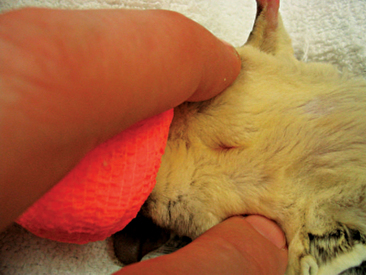
Male sugar gliders have a scent gland on top of their heads; however, owners may report it as a bald spot and think it is the result of the animal’s rubbing its head on the cage, nest box, or directly onto the female. These animals also have a scent gland on their chest (see Figure 11-1). Male sugar gliders, like other marsupials, have unique genitalia. The penis of these animals is bifurcated, and the scrotum is located cranial to the penis (Figure 11-2).
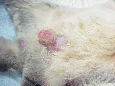


Abdominal masses may indicate that a female glider is pregnant. The symmetry of these masses can reveal the number of young being carried by the female (Figure 11-3). Most pregnant sugar gliders give birth to one or two joeys. Gestation length is a mere 16 days, and once born, the joeys find their way to the pouch at the earliest stages of development. Joeys will remain in the pouch up to 2½ months. Thereafter, the developing young will begin to venture from the pouch, returning primarily to nurse and for protection. The intermittent pouch life will last for a period of 1 to 2 months. The male glider is quite gentle and receptive to these newcomers of the nest box.


Reduced appetite or anorexia may be the result of any number of initiating factors. Stress is one of the more frequent causes of irregular eating patterns affecting sugar gliders. Sources of stress for a sugar glider can include environmental change; lack of a dark, secure hiding place; or absence of a companion. Illness or injury also may cause a decrease in appetite. Abnormal or damaged dentition should be a consideration of the practitioner presented with a glider having a depressed appetite. Cataracts or blindness cause obvious restrictions to eating as well. Dentition and ocular abnormalities have been known to occur in the offspring of obese parents. Blindness likely originates from the inadequate nutrition that was the predisposing factor for the obese condition of the parents rather than the actual obesity itself.


Rear leg paresis or paralysis has been noted within the species. The exact cause(s) remain undetermined, although reduced opportunity for exercise as a result of captivity may play a role. Nutritional deficiencies, such as inadequate protein intake, should be considered, and injections of vitamin B complex, vitamin C, vitamins A and D3, vitamin E and selenium, and calcium may be administered to treat the condition.1


Sugar gliders appearing to miss landings, bounce off walls, or fall frequently may be suffering from one of several disease conditions. Rear limb ataxia, blindness, and obesity can all cause incoordination in gliders. Trauma resulting in long bone fractures, dislocations, or soft tissue trauma can have similar patient presentation.


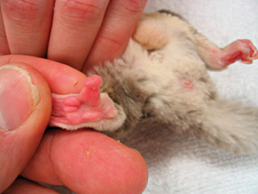
Instability in, or absence of, normal stance should be investigated, commencing with a physical exam. General anesthesia of the patient should be used to ensure a thorough examination of the animal is obtained. Primary muscle bellies should be checked for asymmetry as a result of atrophy or swelling. Digits should be palpated for injury, infection, or foreign material (Figure 11-4). The appendages should be examined for joint symmetry and mobility, flexion and extension. The spinal cord should be palpated for deviation. Radiographs will serve to assist in determining soft tissue or skeletal injury. Appropriate nutrition and husbandry can often help to reduce the incidence of many of these locomotor abnormalities, and as such, client education is essential.


ANESTHESIA AND RESTRAINT


Manual Restraint of Macropods


The type of manual restraint selected for the macropod is dependent upon size. Animals up to 36 kg (80 lb) can be captured manually and then restrained by an experienced handler. The animal is initially caught by grasping the base of the tail with one or two hands. Once a handler gains control of the tail, the animal will tend to bounce upward, and this movement will provide an opportunity for the handler’s arm to be placed around the animal’s chest, just below the forelimbs. The animal can then be lifted up; however, hindlimb movement can injure veterinary personnel or the animal itself. The tail should be angled forward to prevent the ventral surface striking the handler or a hard surface. The goal of macropod restraint is to minimize any opportunity for the animal to gain leverage. The macropod then is tipped head first into a Hessian or denim bag for transport, weighing, examination, blood collection from the lateral tail vein, or the administration of chemical restraint.


Adult macropods of smaller size, 5 to 10 kg (11-22 lb), may be captured using a net on a long pole (1-2 m). The net should be composed of a fine mesh or solid cloth to prevent entanglement or leg injuries. Macropods sensing capture will often run along the fence line of an enclosure. A keeper can take advantage of this flight response by taking a position along the fence line. A space should be maintained between the fence line and the person that is poised to capture the animal. The capture pole with attached net is held parallel to the fence but behind the keeper. Other individuals then herd the selected animal along the fence line toward the net. As the animal passes between the keeper and the fence, the net is brought forward in front of the animal and the animal contained. Personnel involved with capture and restraint should be cautious not to overstress the animal, as stress may result in hyperthermia and subsequent exertional myopathy. If the macropod is not captured on the first attempt, consider postponement of the procedure or darting the animal. Once the animal is netted, basic clinical procedures can be performed. In animals that weigh 36 kg (80 lb) or greater, sedation by remote injection techniques is the safest approach for capture, reducing injury risk to both humans and animal.


Chemical Restraint of Macropods


Chemical restraint of macropods weighing less than 10 kg (22 lb), such as small wallabies, juvenile kangaroos, and wallaroos, may be accomplished with manual restraint and inhalation anesthesia. Anesthetic induction of smaller macropods is best accomplished using 5% isoflurane gas with a 1.5-L flow of oxygen delivered via a face mask. Larger animals require injectable anesthetic agents for chemical immobilization. A variety of these drugs have been used alone or in combination, including pentobarbital, thiopentone, phencyclidine-acepromazine, etorphine, etorphine-acepromazine, etorphine-methotrimeprazine, etorphine-ketamine, droperidol, droperidol-fentanyl, azaperone, alphaxalone-alphadolone, alphachloralose, diazepam, xylazine, ketamine, xylazine-ketamine, tiletamine-zolazepam, and medetomidine-ketamine.


Macropods are especially susceptible to trauma-related injuries and capture myopathy during restraint. The goal of chemical immobilization is a smooth and rapid induction and recovery, thereby reducing the incidence of trauma-related injuries.


Medetomidine-ketamine and tiletamine-zolazepam are two chemical immobilization combinations reported to be particularly effective. Both therapeutic combinations provide a rapid and smooth induction within 10 minutes. The advantage of medetomidine-ketamine over tiletamine-zolazepam is its reversibility with atipamezole. Dosages utilized for these combinations are species dependant. Doses of 40 mg/kg of medetomidine mixed with 4 mg/kg of ketamine administered intramuscularly (IM) have been found to be an effective combination when used on eastern gray kangaroos.1 Ranges of 40 to 70 mg/kg of medetomidine with 4 to 7 mg/kg of ketamine have been found to be effective for red kangaroos, wallaroos, Dorcopsis wallabies (Dorcospsis macleayi), and Goodfellow’s tree kangaroos (Dendrolagus goodfellowi).1 Atipamezole given at a dose 5 times the total mg of administered medetomidine has been found to be effective for reversal in the above-mentioned species.1 A dose of 100 mg/kg of medetomidine and 5 mg/kg ketamine IM has been effective in immobilizing red necked wallabies.1


Tiletamine-zolazepam at a dose of 2 to 8 mg/kg is recommended for chemical immobilization of tree kangaroos.1 In most macropods, however, the general dose range is 3 to 11 mg/kg IM.1


Other chemical combinations administered IM to immobilize macropods include xylazine and ketamine. If either medetomidine and ketamine or tiletamine and ketamine are selected as a combination, a supplemental injectable ketamine anesthetic may be administered as needed. Supplemental ketamine administration can be accomplished by placement of an intravenous (IV) catheter in the lateral tail or lateral saphenous veins.


Propofol offers the benefit of being an ultra–short-acting induction agent. An adverse side effect of propofol administration is an initial transient apnea. To reduce the risk associated with transient apnea, intubation is recommended when this agent is used. The standard mammalian dose of 6 to 8 mg/kg propofol is also the recommended dose for macropods and should be given slowly to effect. Alternatively, an animal can be premedicated with midazolam at 0.2 mg/kg, allowing a lower dose of 5 mg/kg of propofol to be given to effect.


If a surgical plane of anesthesia is desired, supplemental inhalation anesthetic (e.g., isoflurane), in addition to an injectable agent, may need to be supplied. Gas is delivered by a face mask and then followed by endotracheal intubation. The placement of an endotracheal tube is a challenge in macropods because of a narrow mouth opening. To aid in glottis positioning and endotracheal tube placement, the animal should be placed in dorsal recumbency with the head fully extended over the side of a table. By doing so, the glottis is aligned with the angle of the mouth, allowing easier passage of an endotracheal tube. Strips of gauze can be placed through and around the maxilla and mandible as an aid to open the mouth. A laryngoscope with a long blade (15 cm or 6 inches) is recommended to visualize the glottis as a straight, semirigid endotracheal tube is passed into the mouth. Visualization of the intubation procedure can be impeded by the tube and again is best performed utilizing the position described above. If intubation is unsuccessful, an alternative is to pass a smaller diameter tube first, such as a 6-Fr dog urinary polypropylene catheter. This smaller tube can be easily visualized as it is put into the trachea, thus confirming proper placement. The endotracheal tube is then passed over the catheter, which serves as a guide. The tube is secured by first tying a strip of gauze around the tube and then tying the remaining lengths of material behind the base of the skull. Both isoflurane and halothane inhalation anesthetics have been used successfully for macropod anesthetic induction and maintenance.


Sugar Glider Anesthesia


Anesthesia for sugar glider patients is best accomplished with inhalation agents (e.g., isoflurane). A medium- or large-sized mask can be used as an induction chamber, with isoflurane being delivered at a level of 5% and an oxygen flow rate of 1 L/min (Figure 11-5). Once induced, the animal can be transferred to a smaller face mask and maintained at 2% isoflurane (600-800 ml/L oxygen). Placement of an endotracheal tube (e.g., 14-16 gauge intravenous catheter) may be attempted, but it presents an obvious challenge in terms of the animal’s size and the possibility of tube blockage caused by the buildup of respiratory secretions.


Injectable anesthetics are rarely used to anesthetize sugar glider patients because inhalation agents have a much reduced risk and are easily provided. One study reported using tiletamine-zolazepam at doses ranging from 8.4 to 12.8 mg/kg IM with no adverse side effects.1 However, tiletamine-zolazepam at 10 mg/kg has been associated with neurologic problems and death in squirrel gliders (Petaurus norfolcensis), so caution is advised with use of this drug.1 Ketamine has also been used to induce anesthesia in gliders at a recommended dose of 20 mg/kg.1 Acepromazine has been combined with butorphanol and given orally or with ketamine and given subcutaneously (SC) to maintain postoperative sedation and analgesia.


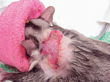
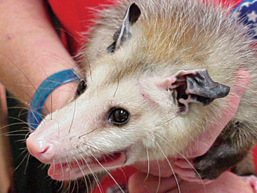
Manual Restraint of Opossums


The private practitioner or wildlife rehabilitator may be presented with a Virginia opossum (Didelphus virginiana) (Figure 11-6). Often, these animals have incurred vehicle or fight injuries, and as a result, attention and care must be taken in their restraint (Figure 11-7). Heavy gloves and towels should be used during the removal of the animal from its enclosure. These animals have particularly sharp teeth and, if not appropriately restrained, will turn quickly to bite. Similarly, opossums retain long claws and may scratch in a threatened situation. A firm grasp of the scruff of the neck and the base of the tail are essential in the movement of the animal. If assistance is available, these restraints can be divided between two individuals. However, if the handler is alone, the animal’s head and neck should be held farthest from the handler and the base of the tail held as the primary means of support in the carriage of the animal. As in other species, the tip of the tail should never be held, as injury may result. Masked induction with 5% isoflurane and 1.5 L/min of oxygen followed by endotracheal intubation and a maintenance rate of 2% to 3% isoflurane and an oxygen flow rate of 1.5 L/min of oxygen is considered sufficient for basic examination, wound care, and diagnostics.
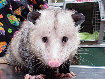


COMMON PRESENTATIONS BY SYSTEM


Respiratory System


Respiratory disease as a primary illness is rarely noted in marsupial species. Rather, respiratory conditions are more often associated with another disease process or as an opportunistic consequence. Similar to that of other companion animal species, diseases associated with the respiratory tract of marsupials may occur as a result of infectious disease (e.g., bacteria, viruses, parasites, fungi, yeast), neoplasia, and trauma. Diagnoses and treatments pursued for these disease processes should follow standard protocols.


FOREIGN BODIES


The foreign bodies most commonly found in the upper respiratory tract of marsupial species are the grass seed and grass awn. Over the course of eating, animals will accidentally inhale seeds, awns, or both. Grass seeds, in particular, will enter the nares of captive, grazing marsupial species, such as kangaroos, wallabies, and wombats. Wombats are susceptible to grass seed inhalation and present with unilateral purulent nasal discharge. Practitioners should be aware that marsupials that have contact with hay or straw, either as a bedding material or in an anesthetic recovery area, inadvertently inhale grass seeds.


Foreign bodies in the nares manifest in symptoms of unilateral purulent nasal discharge, sneezing, and persistent pawing at the nares. The offending foreign body must be visualized before removal. The animal first should be heavily sedated or placed under general anesthesia. An otoscope, rigid arthroscope, or endoscope can then be used to locate the foreign material. Removal is best achieved with alligator forceps. Antibiotics should be prescribed as a prophylaxis against bacterial infection. Clavulanic acid and amoxicillin (Clavamox) can be given to macropods and wombats at a dose of 12.5 mg/kg IM twice a day.1 Alternatively, a Clavamox dose of 25 mg/kg can be given IM or SC once daily. The oral administration of Clavamox in macropods and wombats has been associated with yeast overgrowth in the gastrointestinal tract and subsequent diarrhea; therefore, if Clavamox is prescribed, treatment and patient response should be carefully monitored.


Diagnosis of foreign bodies within the lower respiratory tract of marsupials is infrequent when compared with the upper respiratory system. However, hand-raised marsupials are prone to aspiration of artificial milk formula and accompanying aspiration pneumonia. Aspiration of artificial milk is due most often to a juvenile marsupial receiving milk at too high a flow rate. The source of this high flow rate is usually an artificial teat with an opening that is too large. Squeezing a feed bottle or syringe feeding also can deliver milk in amounts the animal cannot accommodate. Attention to a patient’s body temperature is essential, as hypothermia can predispose an orphaned marsupial to inhalation pneumonia as well. Animals with low body temperatures are sluggish and have a depressed swallow reflex, which can result in the inhalation of artificial milk formula.


VIRAL INFECTIONS


A poxvirus is associated with the development of papillomatous proliferations on the skin of macropods. The areas affected most often by these papillomatous proliferations are the head and extremities. The development of papillomas around the external nares and mouth may interfere with normal respiration. Marsupial species affected by this condition include tammar wallabies, Agile wallabies (Macropus agilis), swamp wallabies (Wallabia bicolor), eastern wallaroos (Macropus robustus), quokkas, red kangaroos (Macropus rufus), eastern gray kangaroos (Macropus giganteus), and western gray kangaroos (Macropus fuliginosus).1 The disease itself is considered self-limiting, but if the growths impair an animal’s ability to eat or breathe, surgical removal is an option.


An orbivirus was reported as causing death in Australian tammar wallabies during the summer months.2 No premonitory signs were evident, and animals were usually found either comatose or dead. Diagnosis was based on history, serologic testing, and postmortem examination. Gross pathologic findings included marked pulmonary congestion and edema, hepatic congestion, and frequently, subcutaneous edema in the hindlimbs.2 No effective treatment is currently available. Vaccine development for this orbivirus is currently in progress.


A herpesvirus has been established as the cause of death in a variety of marsupial species.2 Clinical signs associated with this virus include respiratory rales, conjunctivitis, incoordination, and death.2 Serologic testing is available, but a definitive diagnosis is often made when histopathologic examination of affected liver tissue reveals intranuclear inclusion bodies in hepatocytes. A viable treatment option has yet to be found.


BACTERIAL INFECTIONS


Although bacterial infection of the upper respiratory tract is uncommon in marsupial species, the koala appears to be most susceptible. Bacterial infections reported to cause rhinitis in the koala include Bordetella bronchiseptica, Pseudomonas spp., Corynebacterium spp., Streptococcus spp., Escherichia coli, Micrococcus spp., Staphylococcus spp., Proteus spp., Pasteurella spp., and Enterobacter spp.1 In the koala, Chlamydophila psittaci has been implicated as a cause of nasal discharge.1 Clinical signs of upper respiratory tract disease are not unlike those seen in mammalian species. Bilateral nasal discharge, sneezing, anorexia, and coughing are the more common signs encountered. Nasal discharge can be clear or mucopurulent in appearance. Pharyngitis and regional lymph node enlargement have been seen to occur. If Chlamydophila psittaci is suspected, specific guidelines should be followed for specimen collection, transport, and storage. The practitioner should be aware of the following:1


Swabs of the nasal mucosa can be analyzed with antigen enzyme-linked immunosorbent assay (ELISA) to assist in the diagnosis of chlamydiosis. Antigen ELISA kits are commercially available to practitioners. Antigen detection through direct immunofluorescence can also be performed with a commercial kit, but this technique appears less sensitive than the ELISA.1


Complement fixation tests can be used to detect antibody in serum but are less accurate than other tests available. Testing using polymerase chain reaction (PCR) technology is showing promise in the diagnosis of koala chlamydiosis.


The selection of an antibiotic in the treatment of rhinitis should be based on culture and sensitivity testing. The route of administration can be oral, parenteral, or through nebulization. Saline nebulization, in combination with systemic antibiotic therapy, appears particularly effective.


Effective treatments for chlamydiosis include doxycycline hydrochloride, 5 mg/kg every 7 days IM for 6 weeks; chloramphenicol, 30 mg/kg SC every 12 hours for 7 to 10 days; and oxytetracycline, 5 mg/kg SC every 7 days for a maximum of 4 injections.1 Pseudoephedrine hydrochloride at a dosage of 1 mg/kg every 12 hours can be administered via nebulization or per os (PO) as a decongestant. The mucolytic, bromhexine hydrochloride given at a dosage of 2 mg/kg every 12 hours PO or through nebulization also can help to relieve clinical signs.1 Supplemental feeding should be given to koalas when they are being treated with antibiotics, as antimicrobial effects of the therapeutic agents on the gastrointestinal flora have been associated with significant weight loss.


Bacterial pneumonias are commonly diagnosed in the koala. Organisms that have been identified as a causative agent of koala bacterial pneumonia include Corynebacteria spp., Haemophilus spp., Bordetella bronchiseptica, Pseudomonas aeruginosa, and Streptobacillus moniliformis. The animal may show signs of bilateral purulent nasal discharge, coughing, audible respiratory noises, and anorexia. Recommended diagnostic tests include culture and sensitivity and transtracheal wash. Thoracic radiography will often reveal advancement of disease and give indication to prognosis. Parenteral antibiotics are the treatment of choice and should be selected on the basis of culture and sensitivity results. Nebulization with an appropriate antibiotic can significantly contribute to patient recovery. Mucolytics can be used as a supportive measure and may be added to the nebulization cocktail. A vaccination for Bordetella bronchiseptica is available as a cell-free extract named Canvac-BB (CSL, Ltd., Parkville, Victoria, Australia). The vaccination protocol established for the koala is 2 doses SC 4 weeks apart and then annual boosters thereafter.


Necrobacillosis, commonly known as lumpy jaw, is a tooth root infection frequently encountered in captive macropods. Several factors are believed to foster the development of the disease, including overcrowding and poor husbandry—which lead to excessive fecal contamination, especially around feed stations—and a diet containing soft feed or awns. Bacteroides (Dichelobacter) nodosus and Fusobacterium necrophorum, usually acting synergistically, invade and cause infection. Other bacteria less commonly isolated from tooth root infections include Actinomyces spp. and Corynebacterium spp. Additional opportunistic bacteria will invade as an infection advances and may be the only organisms isolated when cultures are finally obtained. Primary clinical signs associated with necrobacillosis are swelling around the face and jaw, excessive salivation, loss of condition because of difficulty in eating, depression, and lethargy. Proptosis of the globe and squinting may be observed as a result of retrobulbar abscessation. A bacteremia may result, spreading organisms to other areas of the body, including the spleen, liver, stomach, bones, and lungs. Treatment of lumpy jaw is focused on removal of affected teeth and debridement of affected soft tissue. Clindamycin is the antibiotic of choice at a dose of 11 mg/kg PO (capsules or liquid) every 12 hours. Animals find the liquid formulation more palatable than capsules, and an injectable formulation exists but is expensive and restricted to IV use. Duration of treatment should be a minimum of 6 months because of the possibility of disease recurrence. Large doses of penicillin (150 mg/kg procaine penicillin and 112.5 mg/kg benzathine penicillin) every second day for approximately 2 weeks has also been recommended, but efficacy of this treatment course is unknown.1


Prevention of necrobacillosis focuses on providing appropriate diet and husbandry for captive animals. Overcrowding of macropod species should be avoided. Environments should be routinely cleaned to avoid the buildup of fecal material both on the ground and around food sources. A Bacteroides nodosus vaccine has been recommended at the following weights and doses: less than 5 kg, 0.2 ml SC; 5 to 10 kg, 0.25 ml SC; 10 to 20 kg, 0.5 ml SC; and more than 20 kg, 1 ml SC.1 The skin over the ribs is the preferred site for recommended SC vaccine injection. Sterile abscesses have been reported at the vaccine injection sites. The vaccine schedule is 2 doses 4 weeks apart, then once a year.


Mycobacteriosis has been reported as a source of respiratory disease in marsupial species.1 Tuberculosis caused by Mycobacterium bovis has had particular impact on the brushtail possum population in New Zealand. The disease is contracted from possums grazing in pastures frequented by infected animals (e.g., cattle, deer, or other possums). Discharge is emitted from the lymph nodes of these diseased animals and usually serves as the source of pasture contamination. Once a possum is exposed, the organism spreads throughout the body, targeting the lungs and other vital organs. Clinical signs associated with marsupial tuberculosis include dyspnea, depression, anorexia, and weight loss. Treatment of the wild brushtail possum is often not pursued because this animal is considered a pest species in New Zealand. The brushtail possum is viewed as perpetuating Mycobacterium bovis in cattle and deer herds in New Zealand, resulting in significant economic loss.


Mycobacteriosis has had a similar impact on captive tree kangaroos in the United States. It has been reported as a common cause of disease and mortality in three species of tree kangaroos in particular, including Matschie’s (Dendrolagus matschiei), Goodfellow’s (Dendrolagus goodfellowi), and Grizzled (Dendrolagus inustus).1 Mycobacterium avium complex is the cause of more than 90% of infections diagnosed in captive tree kangaroos maintained in the United States. The apparent susceptibility of these animals is attributed to a cellular immune response comparatively lower than what is observed in eutherian mammals or other marsupial species. Clinical signs associated with mycobacteriosis in tree kangaroos include weight loss, dyspnea, lameness, abscesses, neurologic problems, and blindness. Definitive antemortem diagnosis is a challenge with the tree kangaroo, and signalment, history, clinical signs, radiology, and transtracheal washes are included as part of the recommended diagnostic regimen. The treatment protocol for infected tree kangaroos is amikacin, 3 mg/kg IM every 12 hours; rifabutin, 20 mg/kg PO every 24 hours; ethambutol, 20 mg/kg PO every 24 hours, and azithromycin, 20 mg/kg PO every 24 hours.1 The duration of treatment can be quite long, with amikacin usually administered for 10 to 12 weeks and oral medications for 3 years or longer. Unfortunately, treatments have yielded poor results. The mycobacterial organism can often still be detected on cultures of transtracheal washes performed 3 or more years after treatment was initiated. Preventing animals from exposure to the organism is the best approach to controlling this organism. Captive tree kangaroos should not be housed near captive birds because of the risk of transmission posed by Mycobacterium avium complex.


FUNGAL INFECTIONS


One of the more common infections affecting the upper respiratory tract of marsupial species is cryptococcosis. Australian marsupials including brushtail possums, ringtail possums (Pseudocheirus peregrinus), striped possums (Dactylopsila trivirgata), Leadbeater’s possums (Gymnobelideus leadbeateri), tammar wallabies, red-necked wallabies (Macropus rufogriseus), spectacled hare wallabies (Largorchestes conspicillatus), quokkas (Setonix brachyurus), and wombats and echidnas (Tachyglossus aculeatus) have been reported as having the disease.1 The species that appears particularly susceptible, however, is the koala. The causative agent is the yeast-like fungi, Cryptococcus neoformans var gattii. The organism’s close association with river red gums (Eucalyptus camaldulensis), mugga ironbarks (Eucalyptyus sideroxylon), and other red gum species results in extremely high koala exposure rates. Inhalation of the organism from a contaminated environment is believed to be the primary source of exposure. Clinical signs that have been observed in the koala include unilateral or bilateral nasal discharge, sneezing, and anorexia. The usual course of infection starts at the nares and progresses into the sinuses, with secondary lung involvement. Granulomatous formation may occur in the respiratory tract. Once the animal’s respiratory system is infected and there is upper respiratory disease, the organism can infect the central nervous system (CNS) as well. With CNS infection, neurologic signs (e.g., seizures, blindness, behavior changes) may be observed. One of the primary diagnostic tests used to diagnose this disease is the microscopic examination of stained smears collected from the nasal cavity. Diff-Quik staining of the collected sample is easily performed and offers excellent visualization of the yeast-like organisms. India ink may be used as an alternative. Nasal discharge should be cultured, as the organism will grow on Saboraud’s agar at 25° C and 37° C. A latex-cryptococcal antigen test (LCAT) is available to detect soluble cryptococcal capsular polysaccharide antigen in body fluid samples. The LCAT can be a particularly effective test when cryptococcosis is suspected, but the site(s) of infection is (are) unknown. Serial dilutions of serum are combined with a suspension of latex particles coated with hyperimmune rabbit serum, with titers of 1 : 2 or greater considered positive. The strength of a positive response can be an indicator of disease severity and, ultimately, prognosis for treatment response and recovery. The decline in LCAT titer is usually slower when compared with clinical improvement but can still be an effective tool in indicating treatment success and prognosis. Survey radiographs of the nasal cavities, sinuses, and lungs also will assist in localizing areas of infection, and survey computed tomography (CT) scans offer even more precision if available.


The recommended treatment protocol for cryptococcosis is fluconazole, 3 mg/kg PO every 24 hours for 30 to 60 days, with a loading dose of 6 mg/kg.1 Amphotericin B is administered in approximately 50 to 60 ml of Hartmann’s solution or 0.9% NaCl at a dose of 0.5 mg/kg SC every second day for 30 days because of the potential for renal toxicity that exists with amphotericin B.1 Renal function should be checked every 2 weeks. If fluconazole is cost prohibitive, ketoconazole is an acceptable alternative. The dosage offered for ketoconazole is 10 mg/kg PO every 12 hours for 30 to 60 days.1


Aspergillus fumigatus has been implicated as a cause of mycotic pneumonia in the macropod. Itraconazole, fluconazole, and amphotericin B are considered to be effective drugs in the treatment regime. Emmonsia-like fungal spores have been detected in the lungs of common wombats (Vombatus ursinus) and southern hairy-nosed wombats (Lasiorhinus latifrons). As burrowing marsupials, they are suspected to inhale the soil-dwelling fungal spores, which then become lodged deep within the lungs, particularly in the alveolar lumen. Levels of infection were determined as being quite high, yet pulmonary responses were mild. Primary pathologic changes in the lungs were mild interstitial fibroplasias and collections of macrophages and leukocytes. The most severe lesions were assessed to be focal granulomas that contained the occasional giant cell. Overall, infection with Emmonsia-type fungal spores does not appear to produce any truly debilitating respiratory change.


NEOPLASIA


Primary neoplastic change occurs frequently in the koala and dasyurid species. Koalas, in particular, are susceptible to the development of craniofacial tumors. These tumors are limited to the upper respiratory tract, with subsequent distortion of the nasal cavity and paranasal sinuses that produce related clinical signs. Nasal discharge, facial distortion, and epistaxis are strongly indicative of the disease, but radiographs can significantly assist in determining a diagnosis. To distinguish a craniofacial tumor from a cryptococcal granuloma, a biopsy of the mass is recommended. Histologic examination reveals craniofacial tumors to be comprised of a mixture of cartilage and bone. Scattered areas or irregular compartments of hypertrophying chondrocytes are embedded in, or surrounded by, vascular and connective tissue. Different stages of calcification or ossification are also evident. Treatment of these tumors is at best palliative, because recurrence is likely, even with complete surgical removal. Euthanasia should be considered in highly infiltrative or advanced cases.


Another common tumor occurring in the respiratory tract of the koala is mesothelioma. This neoplasm targets the serosal surfaces of body cavities, including the peritoneum and mediastinum. Labored breathing, anorexia, and abdominal distension are the predominant clinical signs associated with mesothelioma growth. Diagnosis is best achieved with thoracocentesis, abdominocentesis, or both, and cytologic testing of the collected sample and radiography. Nodules forming as a result of neoplastic change tend to be fibrous in content and have low cellularity. On histologic exam, mesothelial cells can be expected to predominate, with collagen fibers, ground substance, spindle cells, macrophages, lymphocytes, and neutrophils being present in smaller quantities.

Stay updated, free articles. Join our Telegram channel

Oct 1, 2016 | Posted by in EXOTIC, WILD, ZOO | Comments Off on MARSUPIALS

Full access? Get Clinical Tree

Get Clinical Tree app for offline access