8 Management of Heart Failure
Heart failure is a syndrome in which cardiac dysfunction results in clinical signs associated with venous congestion or reduced cardiac output. It is important to recognize that heart failure results from heart disease and that practically any heart disease can result in heart failure. Though the distinction between low-output (forward) failure and congestive (backward) failure is artificial, heart failure in animals usually takes the form of congestive heart failure (CHF). The clinical signs are primarily the result of the development of abnormally high systemic or, more often, pulmonary venous pressures. Left-sided CHF is typically characterized by the presence of pulmonary congestion and edema. Right-sided CHF is manifested as body cavity effusions, usually ascites, or peripheral edema.
Most heart diseases in dogs are chronic; despite this, it is common for clinical signs to develop suddenly. The reasons for this are varied. Pets are largely sedentary, and, consequently, subclinical heart disease can progress until a minimum of stress or exertion provokes signs of cardiac dysfunction. Additionally, subtle changes in respiratory rate and character are difficult for pet owners to recognize. Regardless, it is common for chronic disorders such as degenerative mitral valve disease and canine dilated cardiomyopathy to result in clinical signs that are apparently sudden in onset and necessitate emergent management. Such a presentation might be best described as decompensation rather than acute heart failure. There are, however, a few examples of truly acute CHF in dogs. Rupture of a first-order mitral valve chorda tendinea can result in sudden elevations of left atrial pressure and acute, left-sided CHF. Similarly, destruction of the aortic or mitral valve by an aggressive infective lesion can also cause acute heart failure. Regardless of the precise pathogenesis, the sudden development of cough or dyspnea related to heart failure is an important veterinary emergency.
Respiratory distress related to pulmonary edema is the most consistent historical finding in severe CHF. Syncope, lack of appetite, cough, and depression may also be part of the animal’s medical history. With the exception of cardiac tamponade, which is discussed in Chapter 7, the onset of right-sided CHF is in general more insidious and less often prompts urgent veterinary evaluation.
The physical examination reflects the effects of diminished cardiac performance and increases in ventricular filling pressures. Often, the disease that has resulted in heart failure is also apparent on physical examination. Tachycardia is a relatively consistent finding. It is important to recognize that the respiratory sinus arrhythmia that is common in healthy dogs results partly from vagal influence. Vagal discharge is inhibited in heart failure and, as a result, the finding of a respiratory-related arrhythmia is virtually incompatible with a diagnosis of overt CHF.
On physical examination, elevated ventricular filling pressures are manifested as dyspnea resulting from pulmonary edema or pleural effusion. Crackles are often audible when pulmonary edema is present, whereas significant pleural effusion may muffle heart and lung sounds.
Auscultatory findings may reflect the disease that is responsible for the heart failure state. Dogs with CHF resulting from degenerative mitral valve disease invariably have a systolic murmur of mitral valve regurgitation; most often, the murmur is loud. Dogs with dilated cardiomyopathy may have more subtle physical examination findings, although careful auscultation often reveals a soft murmur of functional mitral incompetence and/or a gallop rhythm.
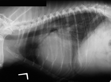
The clinical signs of left-sided heart failure and primary respiratory tract disease are superficially similar. Because aggressive diuretic therapy can be lifesaving in CHF but harmful in the setting of respiratory tract disease, an accurate diagnosis is essential. A noninvasive diagnosis of left-sided CHF can be made radiographically; left atrial enlargement in the presence of pulmonary opacities compatible with edema is diagnostic (Fig. 8-1). Sometimes, the fragile clinical status of the animal is an impediment to careful radiographic examination and the risk-benefit ratio is in favor of empirical therapy. In these cases, a careful assessment of the history and physical examination findings is essential. Before empirical therapy, it is important to determine that a diagnosis of acute CHF is at least plausible.

Figure 8-1 This lateral thoracic radiograph was obtained from a small-breed geriatric dog with congestive heart failure that resulted from degenerative mitral valve disease. The left atrium is enlarged, and there are pulmonary opacities that represent cardiogenic pulmonary edema.
A long history of cough and dyspnea suggests that primary respiratory tract disease is at least partly responsible for the clinical signs, because animals with overt CHF are unlikely to survive for months without treatment.
Heart failure results from heart disease; therefore it is important to consider whether it is likely that the animal has a cardiac disorder that could reasonably result in heart failure. The most common acquired heart diseases that result in left-sided CHF in dogs are dilated cardiomyopathy and degenerative mitral valve disease; the former most commonly affects middle aged, large-breed dogs, whereas the latter affects elderly, small-breed dogs. If CHF is present in an elderly small-breed dog, the murmur is usually loud. Conversely, it is extremely unlikely for an elderly small-breed dog to develop CHF in the absence of a murmur; in these cases, signs such as cough and dyspnea are almost always the result of respiratory tract disease. Dogs with dilated cardiomyopathy may have relatively subtle auscultatory findings. In these instances, a soft murmur or a gallop rhythm may have great clinical importance.
Thoracic radiographs can provide a noninvasive diagnosis of left-sided CHF. When restraint for radiographic studies can be tolerated by the animal, thoracic radiographs guide the therapeutic approach. When available, echocardiography can provide useful information. Echocardiography cannot provide a diagnosis of CHF; however, echocardiography can be used to determine the cause of heart failure. Sometimes, a cursory echocardiographic examination can be performed using minimal restraint and this can be used to determine if the diagnosis of CHF is plausible.
The primary goals in the management of severe CHF are the restoration of normal ventilatory function and sometimes, augmentation of cardiac performance.
The therapy of acute CHF is different from the therapy of chronic CHF in terms of method and intent. Evidence suggests that activation of compensatory neurohumoral mechanisms such as the adrenergic nervous system and the renin-angiotensin-aldosterone system play an important role in the progression of CHF; pharmacologic blunting of these mechanisms has been shown to improve long-term prognosis. For example, drug efficacy studies performed in people indicate that the long-term use of beta blockers has a cardioprotective effect despite the fact these drugs, at least acutely, can have a negative effect on cardiac performance. In contrast, the hemodynamic effect of therapy is of foremost importance in the management of acute CHF and specific therapeutic interventions can be classified based on their effect on the four primary determinants of cardiac output: preload, afterload, contractility, and heart rate.
Preload is the force that distends the ventricle at end-diastole. It is approximated in the living animal by end-diastolic ventricular pressure or volume. Although end-diastolic left ventricular pressure can be estimated through the measurement of the pulmonary capillary wedge pressure, this is not routinely measured in small animals. However, the concept of preload and its pharmacologic manipulation is theoretically useful.
Cardiogenic pulmonary edema results when high mean atrial pressures are communicated to the pulmonary veins and capillaries. Diuretics cause animals to produce large volumes of urine; the administration of a powerful diuretic such as furosemide rapidly decreases intravascular volume and, therefore, preload. The consequent decrease in left atrial pressure alters the Starling forces at the level of the pulmonary capillary and facilitates the reabsorption of edema fluid.
Furosemide is a high-ceiling loop diuretic appropriate for use in the setting of fulminant CHF. Published furosemide doses vary widely and to a large extent the dose and dosing interval are dictated by clinical response. Intravenous doses in the range of 2 to 7 mg/kg in dogs are reasonable when faced with animals with fulminant CHF. The intravenous route is generally preferred when it can be obtained without imposing undue stress on the animal. Other diuretics such as the thiazides, spironolactone, and amiloride may have a role in the management of animals with chronic CHF; however, they have a weaker diuretic effect than does furosemide and see little use in the treatment of fulminant CHF.
Nitrates such as nitroglycerin (NG) may have beneficial effects in severe CHF. These drugs cause dilation of capacitance veins and specific arterial beds through the release of nitric oxide. The clinical efficacy of NG in small animals is uncertain; however, based on what is known of its pharmacology, the administration of NG would be expected to decrease thoracic blood volume and therefore preload, limiting the accumulation of pulmonary edema.
The transdermal formulation of NG is used most often. Transdermal NG is available as a cream and as a controlled-dose adhesive patch. The former allows somewhat greater flexibility of dosing. In dogs, the dose is adjusted based on body weight; smaller dogs may receive ¼ inch of NG and large dogs as much as 1 inch. NG is commonly applied to the interior of the pinnae, although more central sites such as the inguinal area might lead to more rapid and reliable absorption.
Although it is seldom used, phlebotomy is an alternative means of rapidly decreasing preload. It has the obvious disadvantage that it results not only in the loss of intravascular fluid, but also in a loss of blood cells and proteins.
Preload is related to cardiac performance through the Frank-Starling law of the heart; a decrease in preload diminishes the force of ventricular contraction and therefore decreases stroke volume. As a result, preload reduction generally results in a decrease in cardiac output. However, a few factors likely mitigate this effect in some cases.
Animals with systolic heart failure, for example, from mitral valve regurgitation or dilated cardiomyopathy have large ventricles and, potentially, a “flat” Frank-Starling relationship. That being the case, preload can be reduced until pulmonary vein pressures are low enough to prevent edema formation with little effect on stroke volume. When mitral valve regurgitation is the main factor that limits stroke volume, a reduction in preload can decrease mitral annular dilation, reduce valvular regurgitation, and conceivably increase stroke volume.
In general, preload reduction can be expected to have a negative effect on cardiac performance but this effect is buffered by a large ventricular volume. Animals that have dyspnea because of respiratory disease are likely to have normal or decreased ventricular filling pressures and therefore tolerate aggressive diuresis poorly.
Resistance is the quantity that determines the amount of blood that flows through a vascular bed when subject to a given pressure. In a sense, resistance is a measure of how difficult it is to force blood through the vessels. Resistance is dependent on a number of factors, but the most important of these is vessel diameter because this factor is subject to physiologic influences and manipulation by vasodilating drugs.
Vascular resistance is an important determinant of blood pressure and therefore afterload. However, afterload is best approximated by wall stress, which is related not only to blood pressure, but also to ventricular diameter and wall thickness. If ventricular dilation is not offset by hypertrophy, afterload is increased even if blood pressure is normal. This is the case in animals with dilated cardiomyopathy; the dilated, thin-walled ventricle functions at a mechanical disadvantage because afterload is high even if blood pressure is normal.
In CHF, increases in adrenergic tone and vasoactive hormones such as angiotensin II result in vasoconstriction. In the short term, this has the favorable effect of maintaining systemic blood pressure when cardiac output is low. However, in animals with dilated, thin-walled ventricles, systolic wall stress (afterload) is elevated; this increase in afterload comes at the price of an increase in myocardial oxygen demand. When systolic myocardial failure is present, there is a mismatch between contractility and afterload. Judicious vasodilation reduces afterload, which permits an increase in stroke volume. When mitral valve regurgitation is the cause of CHF, there is an analogous mismatch between vascular resistance and the resistance imposed by the regurgitant orifice; the compensatory increase in vascular resistance is a factor that limits stroke volume.
Vasodilators are not indicated in systolic failure because systemic hypertension is present; rather, they are effective because they decrease vascular resistance and allow stroke volume to increase. The favorable effect of vasodilation is one of degree; excessive vasodilation results in hypotension in animals with systolic failure as it does in normal individuals.
Nitroprusside is a balanced vasodilator that has a pronounced effect on the systemic arterioles. Metabolism of nitroprusside is rapid and results in the release of cyanide and nitric oxide; the latter possesses vasodilatory properties. The agent is infused intravenously at a rate of 1 to 10 μg/kg/min. Nitroprusside is a potent vasodilator and should be used only in carefully monitored circumstances. Measurement of systemic blood pressure is recommended and the dose should be titrated based on serial blood pressure measurements and indices of peripheral perfusion. Cyanide toxicosis is a potential adverse effect associated with the use of this drug and it is suggested that the use of nitroprusside be limited to periods of less than 48 hours.
