Chapter 41 Malassezia Dermatitis
Malassezia dermatitis (MD) is a superficial fungal (yeast) infection occurring on and within the stratum corneum of the epidermis of many mammalian species. There are several different species of Malassezia yeast recognized, and various animals may serve as the natural hosts for specific species of the yeast. For example, most domestic carnivores harbor Malassezia pachydermatis as part of their natural cutaneous microflora, while human beings primarily harbor Malassezia furfur. As commensal organisms, Malassezia yeast colonize the skin in very low numbers. Overt infection is defined by increased numbers of the yeast on the skin surface in conjunction with inflammation. In dogs with atopic dermatitis (AD), M. pachydermatis may be recognized by the immune system as an allergen, in which case a highly inflammatory and pruritic response can be mounted to relatively low numbers of yeast organisms, blurring the line between “colonization” and “infection.” The role of M. pachydermatis in feline dermatitis is less well defined, although it is a known commensal of feline skin as well.
PATHOGENESIS
Canine Malassezia Dermatitis
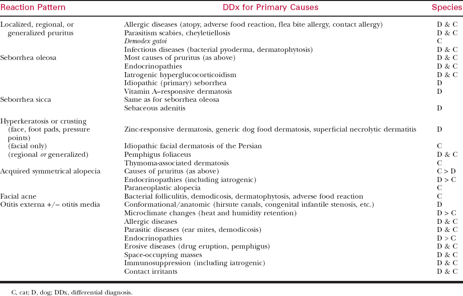
Table 41-1 COMMON DIFFERENTIAL DIAGNOSES FOR SKIN DISORDERS THAT MAY BE ASSOCIATED WITH SECONDARY MALASSEZIA DERMATITIS
Canine Malassezia Otitis
Feline Malassezia Dermatitis and Otitis
CLINICAL SIGNS
Canine Malassezia Dermatitis
Stay updated, free articles. Join our Telegram channel

Full access? Get Clinical Tree


