58 Loss of balance
INTRODUCTION
Sudden onset of idiopathic peripheral vestibular disease is very common in the older dog. Vomiting may immediately precede the loss of balance. The animal leans and falls to one side and as a result is usually recumbent. The dog appears to struggle to remain upright and pays little attention to the owner while doing so. This dramatic presentation is often mistaken for a seizure, especially if the animal has also urinated. Examination in the acute phase always detects a head tilt and spontaneous jerk nystagmus. The head and eyebrows may also twitch in synchrony with the nystagmus. The animal is alert. Postural testing to determine proprioception and strength is almost impossible in the first 24–48 hr due to the extreme ataxia. Two people are required to support the larger dog on a non-slip surface while hopping and ‘knuckling’ is attempted. Neurological signs correspond to a unilateral peripheral vestibular lesion. The age and speed of onset are highly suggestive of the diagnosis.
Signs do not worsen with time. Improvement is slow and is expected within a few days. Ataxia resolves first, followed by cessation of the spontaneous nystagmus. Ambulation without assistance may take 2–3 weeks but the prognosis for return to normal function is excellent. A residual head tilt may persist for months. Recurrence has been reported. Treatment is purely supportive.
Idiopathic peripheral vestibular disease also occurs in cats of any age, not specifically the geriatric animal. Onset and improvement follow a similar pattern to that of the dog. In a group of 75 affected cats, approximately half presented with a head tilt, ataxia and no spontaneous nystagmus. Six cats had bilateral peripheral vestibular disease.
CASE HISTORY
A left head tilt and balance loss began 1 month ago and had not improved. The dog was unsteady when jumping off furniture and had fallen over.
The dog was unable to localize the direction of sound. Mild otitis externa had been an intermittent problem over the years.
CLINICAL EXAMINATION
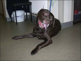
The dog was alert, ambulatory and had a normal gait. Hopping and proprioception were also normal. A spontaneous rotatory nystagmus with the fast phase to the right, slight positional ventral strabismus of the left eye and a left head tilt were present (Fig. 58.1). The left upper lip appeared to droop, owing to the left head tilt. Facial muscle function was normal bilaterally. Facial sensation was present bilaterally.
NEUROANATOMICAL DIAGNOSIS
The lesion was localized to the left peripheral vestibular apparatus – the inner ear to affect both the cochlear duct and semicircular canals given the inability to localize sound as well as the loss of balance or the surface of the brainstem where the left CN VIII enters (Table 58.1).
Table 58.1 Differentiation of central and peripheral vestibular disease
Peripheral | Central | |
---|---|---|
Nystagmus (type) |
![]() Stay updated, free articles. Join our Telegram channel![]() Full access? Get Clinical Tree![]() ![]() ![]() |