44 LMN paresis and paralysis
Acquired myasthenia gravis
INITIAL PRESENTATION
Dysphagia, regurgitation, retching, dysphonia, ptyalism, inability to blink, cervical ventroflexion (cat), tremors, stiffness progressing to generalized weakness with exercise, hindlimb weakness, recumbency and eventually respiratory failure.
INTRODUCTION
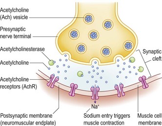
In the normal animal, the arrival of an action potential in a nerve terminus causes the voltage-gated calcium channels to open. The calcium influx induces a coordinated release of acetylcholine (Ach) into the synaptic cleft. The Ach binds to the Ach receptor on the skeletal muscle endplate, opening the membrane to sodium ions which depolarizes the post-synaptic membrane. Muscle contraction follows. The enzyme acetylcholinesterase rapidly removes the Ach from the synaptic cleft, allowing depolarization to recur (Fig. 44.1).
Acquired myasthenia gravis is an immune-mediated destruction of the acetylcholine receptors of the skeletal muscle endplate that interferes with neuromuscular transmission causing weakness.
CASE HISTORY
A husky, harsh-sounding bark and drooling were noted 5 days prior to referral. The dog began to retch and brought up a clear sticky phlegm the following day. Exercise intolerance quickly developed, limiting the dog to walking only a few yards before going stiff, trembling and sitting down. The dog sat rapidly while attempting to squat when urinating or defecating.
The next day the dog collapsed and was presented for examination.
CLINICAL EXAMINATION
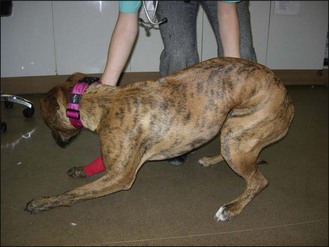
The dog was alert and could stand without assistance (Fig. 44.2). She quickly developed an arched back, all her limbs extended, and she quickly sat and then lay down. Proprioception was normal. Hopping was normal in those brief periods of standing. Spinal reflexes were normal to increased. Muscle tone was normal. No atrophy or muscle pain was noted. Pain perception appeared normal in all limbs. The blink reflex was weak bilaterally with the lids only partially closing. Facial sensation was normal.
DIFFERENTIAL DIAGNOSIS OF ACUTE LMN SIGNS
Immune-mediated myasthenia gravis
Signs reflect skeletal muscle weakness and may be focal (facial, pharyngeal, laryngeal, oesophageal) or generalized (focal signs + appendicular/axial weakness). The most common initial signs in dogs are generalized weakness with megaoesophagus or megaoesophagus alone. In one study, 13% of seropositive dogs had generalized weakness without oesophageal or pharyngeal weakness. Cats chiefly present with generalized weakness and a decreased palpebral reflex. Extraocular muscle weakness is not reported in the small animal. Acquired myasthenia gravis is more common in the dog than the cat.
There is a higher prevalence in certain dog and cat breeds. A sudden onset, rapidly worsening generalized form, described as acute and fulminating, occurs in approximately 15% of cats and dogs with myasthenia gravis.
Signs may worsen with cold weather, oestrus cycles, pregnancy, concurrent infection and vaccination.
The age of onset ranges from 7 weeks to 15 years of age. Two peaks occur: at approximately 2–3 years and 9–10 years.
Tick paralysis
Envenomation by the Ixodes (Australia) or Dermacentor (USA) ticks causes a sudden onset, rapidly ascending flaccid paralysis by preventing release of Ach from the nerve terminus. Cats appear resistant to the salivary neurotoxin of Dermacentor spp. Clinical cases of tick paralysis in Australia coincide with the larger tick population in the spring and summer. Dogs and cats are affected most commonly in late winter and early spring prior to developing humoral immunity.
Ascending flaccid paralysis occurs 5–9 days after the attachment of the Dermacentor tick, progressing to tetraplegia within 3 days. Spinal reflexes are absent (patellar reflex lost before withdrawal reflex). A bark reduced in volume and changed in character, facial weakness and jaw weakness are found.
Signs commence 5–7 days after the attachment of the Ixodes tick, depending on the rate of feeding rather than the absolute number of ticks. High ambient temperatures shorten the course of the disease while in cold weather, signs may not commence until 2 weeks after tick attachment. Ascending flaccid paralysis progresses from hind to forelimbs within a few hours, leaving the animal in lateral recumbency. Spinal stretch reflexes such as the patellar reflex are quickly lost, followed by the withdrawal reflex which weakens as the disease progresses. The gag reflex is absent leading to pseudoptyalism. Voice change and regurgitation are common and early findings. Hypothermia develops. Centrally mediated sympathetic stimulation produces peripheral vasoconstriction and systemic hypertension with secondary pulmonary oedema. Tachycardia or reflex bradycardia and pupil dilation also occur. The pupillary light reflex eventually fails. Respiratory rate falls but tidal volume remains unchanged. Respiration is laboured, with a grunt on expiration from closure of the vocal folds. Cats have a similar presentation with initial signs of agitation, coughing, retching, a changed voice and prominent mydriasis.
Some dogs affected by Ixodes ticks have focal weakness of laryngeal, pharyngeal and oesophageal muscles without appendicular weakness. Megaoesophagus with or without dysphagia may be the sole presenting sign.
Diagnosis is aided by clinical improvement seen after tick removal. Engorged ticks may have fallen off by the time the animal is presented.
Envenomation
Snakes, spiders, lizards, hornets, and worms found in Greenland shark meat also interfere with neuromuscular transmission. Familiarity with the local toxic fauna is recommended.
Rabies
Rabies is transmitted by bites from an infected animal. The time between the bite and demonstration of neurological signs can vary from 1 week to 6 months. Behaviour change (pica, nervousness and fearfulness), ataxia and paresis (limbs, jaw and tongue) are reported, with death occurring within 7–10 days. The chief point to remember about rabies is that it has a highly variable clinical presentation.
Botulism
Ingestion of the preformed type C. botulinum toxin in carrion and rotten food produces a sudden onset of ascending flaccid paralysis, the severity of which is linked to the amount of toxin ingested. The toxin prevents release of Ach from the nerve terminal. The onset of signs occurs within 0.5–6 days after ingestion. Hindlimb followed by forelimb paralysis occurs. Spinal reflexes are reduced or absent. Facial weakness, decreased jaw tone, reduced gag reflex and megaoesophagus are prominent. Autonomic manifestations may occur: mydriasis decreased PLR, decreased lacrimation, urine retention and constipation.
Diagnosis is made from the clinical signs and a history of eating carrion or rotten meat such as found in parks or fed in hunt kennels. It is an uncommon cause of paralysis. Recovery occurs within 1–3 weeks. Death from respiratory paralysis or aspiration pneumonia is possible.
Acute idiopathic polyradiculoneuritis
Inflammation of the ventral nerve roots occurs in dogs, with or without a raccoon bite or scratch. Ascending flaccid paralysis develops spontaneously or 7–12 days after raccoon exposure. Vaccination has been associated with acute polyradiculoneuritis in a young dog. Progression within 3–10 days to non-ambulatory tetraparesis or tetraplegia is accompanied by loss of voice and facial weakness. Affected dogs are able to eat if given access to food. Respiratory paralysis is more likely with rapidly worsening tetraparesis. The perineal reflex is present while other spinal reflexes are absent. Neurogenic atrophy is a feature, dysphagia is not. Hyperesthesia to innocuous touch has been reported.
Diagnosis is aided by finding an elevated level of total protein in the lumbar spinal fluid, reflecting the position of the nerve roots within the subarachnoid space (Table 44.1). Widespread denervation potentials are recorded on electromyography (EMG).
Polymyositis
Myopathies are characterized by exercise intolerance, a stiff, stilted gait, lameness alternating between limbs, lameness worsening with exercise (cf. orthopaedic disease), muscle swelling or atrophy and pain. Weakness may occur but proprioception is normal and spinal reflexes are generally intact. Dysphagia, regurgitation, masticatory muscle atrophy and a weak bark also occur. AST, ALT and CK are not uniformly elevated in myopathies. If raised, their decline may be used to monitor any response to treatment.
Autoimmune polymyositis
is an autoimmune disease of unknown aetiology mostly affecting adult large breed dogs of either sex. Infectious myopathies caused by protozoa or tick-borne pathogens are rare but efforts must be made to rule them out with serology. Equally, drug-induced myopathies should be considered when taking a history. Paraneoplastic myopathies are rare. A generalized immune mediated disease may present as polymyositis.
In this case preservation of spinal reflexes, muscle tone and proprioception was suggestive of myasthenia gravis. Generalized weakness from cardiac insufficiency or hypoadrenocorticism could produce exercise intolerance and collapse.

Stay updated, free articles. Join our Telegram channel

Full access? Get Clinical Tree

