44 LIVER CYTOLOGY
2 What is the cytologic appearance of normal hepatocytes?
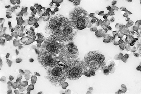
Normal hepatocytes will be present in small to large clusters or groups as well as individually in the smear. Hepatocytes are large epithelial cells with abundant, light-blue cytoplasm that is generally oval to polyhedral in shape. The cytoplasm often appears grainy because of the contrast between high numbers of blue-staining ribosomes and clear to lightly pink–staining organelles. Hepatocytes generally have round, uniform nuclei with moderately coarse chromatin and one or multiple small nucleoli (Figure 44-1).
3 What is the cytologic appearance of normal biliary tract epithelial cells?
Biliary tract epithelial cells are generally present in low numbers, with only a few small groups or sheets of 15 to 20 cells observed. These cells are usually cuboidal to low columnar cells. They tend to have a round nucleus that is often centrally placed in a small amount of light-blue cytoplasm. Nucleoli are not visible.
5 How is bile pigment recognized cytologically?
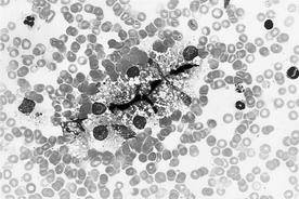
Bile pigment appears as bright- to dark-green granules within the cytoplasm of hepatocytes or as dark-green to black bile casts between hepatocytes (in bile canaliculi) (Figure 44-2). Increased amounts of bile pigment indicates cholestasis, however, the cholestatic process may not be severe enough to cause hyperbilirubinemia or jaundice. Some bile pigment may be present in the cytoplasm of normal hepatocytes. The presence of bile casts indicates cholestasis. Pigment can be confirmed as bile pigment by staining some smears with Hall’s stain, which causes bile pigments to stain green. Staining to confirm bile pigment is seldom done.
8 How is hepatic copper accumulation recognized cytologically, and in what dog breeds is it most often found?
Stay updated, free articles. Join our Telegram channel

Full access? Get Clinical Tree


